The HDAC3 Antibody (CAB2139) is a high-quality antibody developed for reliable detection and analysis of target proteins. This antibody, produced in rabbits, shows high specificity for human samples and has been validated for use in Western blot applications. It binds specifically to the HDAC3 protein, allowing for accurate detection and analysis in a variety of cell types.HDAC3, a member of the histone deacetylase family, is known to play a pivotal role in chromatin remodeling and gene expression. Dysregulation of HDAC3 has been linked to various diseases, including cancer, neurodegenerative disorders, and metabolic syndromes.
This antibody is validated for use in WB, IHC-P, IF/ICC, ELISA applications and has demonstrated reactivity against Human, Mouse, Rat samples.
Product Name:
HDAC3 Antibody
SKU:
CAB2139
Size:
20μL, 100μL
Reactivity:
Human, Mouse, Rat
Conjugate:
Unconjugated
Immunogen:
Recombinant protein (or fragment).This information is considered to be commercially sensitive.
Recommended starting concentration is 1 μg/mL. Please optimize the concentration based on your specific assay requirements.
Synonyms:
HD3, RPD3, KDAC3, RPD3-2, HDAC3
Positive Sample:
HeLa, MCF7, NIH/3T3, C6
Cellular Localization:
Cytoplasm, Nucleus, Cytosol.
Calculated MW:
49kDa
Observed MW:
49kDa
Histones play a critical role in transcriptional regulation, cell cycle progression, and developmental events. Histone acetylation/deacetylation alters chromosome structure and affects transcription factor access to DNA. The protein encoded by this gene belongs to the histone deacetylase/acuc/apha family. It has histone deacetylase activity and represses transcription when tethered to a promoter. It may participate in the regulation of transcription through its binding with the zinc-finger transcription factor YY1. This protein can also down-regulate p53 function and thus modulate cell growth and apoptosis. This gene is regarded as a potential tumor suppressor gene.
Purification Method
Affinity purification
Gene ID
8841
RRID
AB_2764158
Buffer Information
Store at -20℃. Avoid freeze / thaw cycles. Buffer: PBS containing 50% glycerol, preserved with proclin300 or sodium azide, pH 7.3.
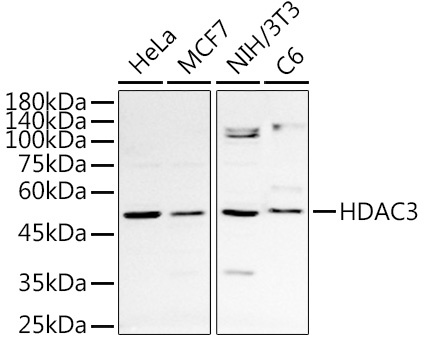
Western blot analysis of various lysates using HDAC3 Rabbit pAb (CAB2139) at 1:1000 dilution. Secondary antibody: HRP-conjugated Goat anti-Rabbit IgG (H+L) (CABS014) at 1:10000 dilution. Lysates/proteins: 25μg per lane. Blocking buffer: 3% nonfat dry milk in TBST. Detection: ECL Basic Kit (AbGn00020). Exposure time: 90s.
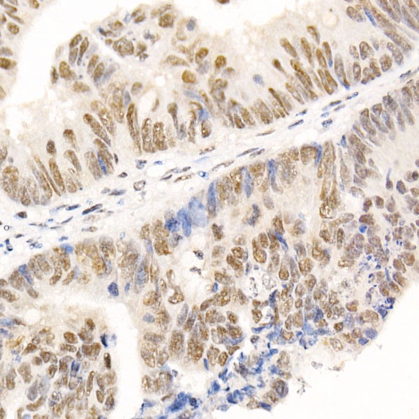
Immunohistochemistry analysis of paraffin-embedded Human colon carcinoma using HDAC3 Rabbit pAb (CAB2139) at dilution of 1:50 (40x lens). High pressure antigen retrieval performed with 0.01M Citrate buffer (pH 6.0) prior to IHC staining.
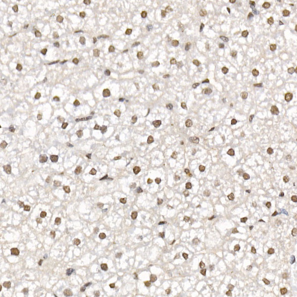
Immunohistochemistry analysis of paraffin-embedded Mouse liver using HDAC3 Rabbit pAb (CAB2139) at dilution of 1:50 (40x lens). High pressure antigen retrieval performed with 0.01M Citrate buffer (pH 6.0) prior to IHC staining.
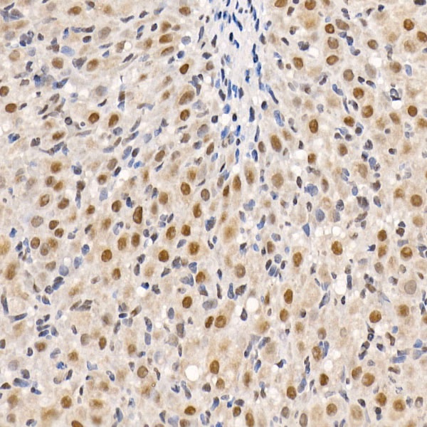
Immunohistochemistry analysis of paraffin-embedded Rat ovary using HDAC3 Rabbit pAb (CAB2139) at dilution of 1:50 (40x lens). High pressure antigen retrieval performed with 0.01M Citrate buffer (pH 6.0) prior to IHC staining.
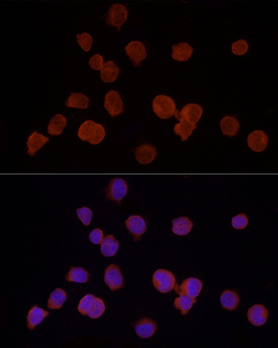
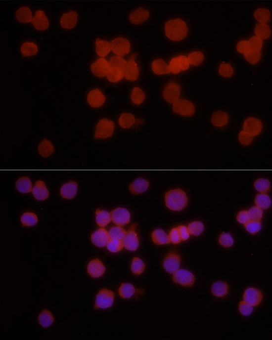
Immunofluorescence analysis of K-562 cells using HDAC3 Rabbit pAb (CAB2139) at dilution of 1:50 (40x lens). Secondary antibody: Cy3-conjugated Goat anti-Rabbit IgG (H+L) (CABS007) at 1:500 dilution. Blue: DAPI for nuclear staining.
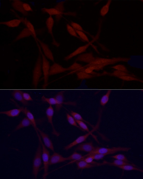
Immunofluorescence analysis of PC-12 cells using HDAC3 Rabbit pAb (CAB2139) at dilution of 1:50 (40x lens). Secondary antibody: Cy3-conjugated Goat anti-Rabbit IgG (H+L) (CABS007) at 1:500 dilution. Blue: DAPI for nuclear staining.
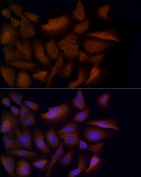
Immunofluorescence analysis of U2OS cells using HDAC3 Rabbit pAb (CAB2139) at dilution of 1:50 (40x lens). Secondary antibody: Cy3-conjugated Goat anti-Rabbit IgG (H+L) (CABS007) at 1:500 dilution. Blue: DAPI for nuclear staining.
Immunofluorescence analysis of K-562 cells using HDAC3 Rabbit pAb (CAB2139) at dilution of 1:50 (40x lens). Secondary antibody: Cy3-conjugated Goat anti-Rabbit IgG (H+L) (CABS007) at 1:500 dilution. Blue: DAPI for nuclear staining.
Immunofluorescence analysis of PC-12 cells using HDAC3 Rabbit pAb (CAB2139) at dilution of 1:50 (40x lens). Secondary antibody: Cy3-conjugated Goat anti-Rabbit IgG (H+L) (CABS007) at 1:500 dilution. Blue: DAPI for nuclear staining.