The HDGF Antibody (CAB13654) is a high-quality antibody developed for reliable detection and analysis of target proteins. This gene encodes a member of the hepatoma-derived growth factor family. The encoded protein has mitogenic and DNA-binding activity and may play a role in cellular proliferation and differentiation. High levels of expression of this gene enhance the growth of many tumors. This gene was thought initially to be located on chromosome X; however, that location has been determined to correspond to a related pseudogene. Alternatively spliced transcript variants encoding distinct isoforms have been described.
This antibody is validated for use in WB, IHC-P, IF/ICC, ELISA applications and has demonstrated reactivity against Human samples.
Product Name:
HDGF Antibody
SKU:
CAB13654
Size:
100μL, 20μL
Reactivity:
Human
Conjugate:
Unconjugated
Immunogen:
Recombinant protein (or fragment).This information is considered to be commercially sensitive.
Tested Applications:
WBIHC-PIF/ICCELISA
Recommended Dilution:
WB
1:500 - 1:2000
IHC-P
1:50 - 1:200
IF/ICC
1:50 - 1:200
ELISA
Recommended starting concentration is 1 μg/mL. Please optimize the concentration based on your specific assay requirements.
Synonyms:
HMG1L2, HDGF
Positive Sample:
22Rv1, U-251MG, MCF7
Cellular Localization:
Cytoplasm, Nucleus.
Calculated MW:
27kDa
Observed MW:
37kDa
This gene encodes a member of the hepatoma-derived growth factor family. The encoded protein has mitogenic and DNA-binding activity and may play a role in cellular proliferation and differentiation. High levels of expression of this gene enhance the growth of many tumors. This gene was thought initially to be located on chromosome X; however, that location has been determined to correspond to a related pseudogene. Alternatively spliced transcript variants encoding distinct isoforms have been described.
Purification Method
Affinity purification
Gene ID
3068
RRID
AB_2760516
Buffer Information
Store at -20℃. Avoid freeze / thaw cycles. Buffer: PBS containing 50% glycerol, preserved with proclin300 or sodium azide, pH 7.3.
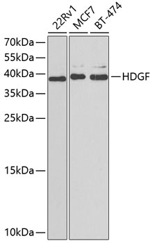
Western blot analysis of various lysates using HDGF Rabbit pAb (CAB13654) at 1:1000 dilution. Secondary antibody: HRP-conjugated Goat anti-Rabbit IgG (H+L) (AS014) at 1:10000 dilution. Lysates/proteins: 25μg per lane. Blocking buffer: 3% nonfat dry milk in TBST. Detection: ECL Basic Kit (AbGn00020). Exposure time: 90s.
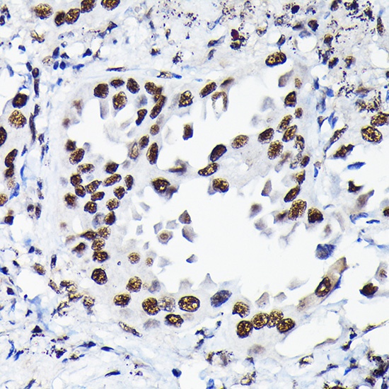
Immunohistochemistry analysis of paraffin-embedded Human lung cancer using HDGF Rabbit pAb (CAB13654) at dilution of 1:50 (40x lens). High pressure antigen retrieval performed with 0.01M Citrate buffer (pH 6.0) prior to IHC staining.
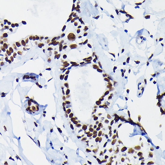
Immunohistochemistry analysis of paraffin-embedded Human breast cancer using HDGF Rabbit pAb (CAB13654) at dilution of 1:50 (40x lens). High pressure antigen retrieval performed with 0.01M Citrate buffer (pH 6.0) prior to IHC staining.