The HDGFRP3 Antibody (CAB8815) is a high-quality antibody developed for reliable detection and analysis of target proteins. This antibody, raised in rabbits, is highly specific and reactive with human samples, making it ideal for experiments in cancer biology and oncology. It has been validated for use in Western blot applications, allowing for the detection and analysis of HDGFRP3 in various cell types.HDGFRP3, also known as Hepatoma-Derived Growth Factor-Related Protein 3, is a potential therapeutic target in cancer treatment due to its involvement in promoting cell proliferation and metastasis.
This antibody is validated for use in WB, IHC-P, ELISA applications and has demonstrated reactivity against Human, Mouse, Rat samples.
Product Name:
HDGFRP3 Antibody
SKU:
CAB8815
Size:
20μL, 100μL
Reactivity:
Human, Mouse, Rat
Conjugate:
Unconjugated
Immunogen:
Recombinant protein (or fragment).This information is considered to be commercially sensitive.
Recommended starting concentration is 1 μg/mL. Please optimize the concentration based on your specific assay requirements.
Synonyms:
HDGF2, HRP-3, HDGF-2, CGI-142, HDGFRP3
Positive Sample:
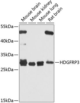
Mouse brain, Mouse kidney, Mouse lung, Rat brain
Cellular Localization:
Nucleus.
Calculated MW:
22kDa
Observed MW:
28kDa
Predicted to enable double-stranded DNA binding activity; microtubule binding activity; and transcription coregulator activity. Predicted to be involved in several processes, including microtubule polymerization; negative regulation of microtubule depolymerization; and neuron projection development. Located in nucleoplasm.
Purification Method
Affinity purification
Gene ID
50810
RRID
AB_2769755
Buffer Information
Store at -20℃. Avoid freeze / thaw cycles. Buffer: PBS containing 50% glycerol, preserved with proclin300 or sodium azide, pH 7.3.
Western blot analysis of various lysates using HDGFRP3 Rabbit pAb (CAB8815) at 1:1000 dilution. Secondary antibody: HRP-conjugated Goat anti-Rabbit IgG (H+L) (CABS014) at 1:10000 dilution. Lysates/proteins: 25μg per lane. Blocking buffer: 3% nonfat dry milk in TBST. Detection: ECL Basic Kit (AbGn00020). Exposure time: 10s.
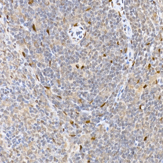
Immunohistochemistry analysis of paraffin-embedded Rat spleen using HDGFRP3 Rabbit pAb (CAB8815) at dilution of 1:100 (40x lens). High pressure antigen retrieval performed with 0.01M Citrate buffer (pH 6.0) prior to IHC staining.