The HEPACAM Antibody (CAB11589) is a high-quality antibody developed for reliable detection and analysis of target proteins. This antibody, produced in rabbits, demonstrates high reactivity with human samples and is validated for use in Western blot applications. By binding specifically to the HepaCAM protein, this antibody allows for accurate detection and analysis in various cell types, making it an essential resource for studies in cancer biology and liver disease research.HepaCAM plays a crucial role in cell adhesion and migration, making it a significant factor in tumor progression and metastasis, particularly in liver cancer.
This antibody is validated for use in WB, ELISA applications and has demonstrated reactivity against Human, Mouse, Rat samples.
Product Name:
HEPACAM Antibody
SKU:
CAB11589
Size:
20μL, 100μL
Reactivity:
Human, Mouse, Rat
Conjugate:
Unconjugated
Immunogen:
Recombinant protein (or fragment).This information is considered to be commercially sensitive.
Recommended starting concentration is 1 μg/mL. Please optimize the concentration based on your specific assay requirements.
Synonyms:
HEPN1, MLC2A, MLC2B, GlialCAM, HEPACAM
Positive Sample:
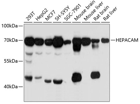
293T, HepG2, MCF7, SH-SY5Y, SGC-7901, Mouse brain, Mouse liver, Rat brain, Rat liver
Cellular Localization:
Cytoplasm, Cytoplasmic Side, Membrane, Single-Pass Type I Membrane Protein.
Calculated MW:
46kDa
Observed MW:
70kDa
The protein encoded by this gene is a single-pass type I membrane protein that localizes to the cytoplasmic side of the cell membrane. The encoded protein acts as a homodimer and is involved in cell motility and cell-matrix interactions. The expression of this gene is downregulated or undetectable in many cancer cell lines, so this may be a tumor suppressor gene.
Purification Method
Affinity purification
Gene ID
220296
RRID
AB_2758617
Buffer Information
Store at -20℃. Avoid freeze / thaw cycles. Buffer: PBS with 0.01% thimerosal,50% glycerol,pH7.3.
Western blot analysis of various lysates using HEPACAM Rabbit pAb (CAB11589) at 1:3000 dilution. Secondary antibody: HRP-conjugated Goat anti-Rabbit IgG (H+L) (CABS014) at 1:10000 dilution. Lysates/proteins: 25μg per lane. Blocking buffer: 3% nonfat dry milk in TBST. Detection: ECL Basic Kit (AbGn00020). Exposure time: 10s.