The HEXA Antibody (CAB5646) is a high-quality antibody developed for reliable detection and analysis of target proteins. This antibody, produced in rabbits, is highly specific for human samples and has been validated for use in Western blot applications. It binds specifically to the Hexa protein, allowing for accurate detection and analysis in a variety of cell types.Hexa is essential for the activity of the enzyme beta-hexosaminidase A, which plays a key role in breaking down certain complex carbohydrates in lysosomes. Mutations in the Hexa gene can lead to lysosomal storage diseases such as Tay-Sachs disease.
This antibody is validated for use in WB, IHC-P, ELISA, IF-P applications and has demonstrated reactivity against Human, Mouse, Rat samples.
Product Name:
HEXA Antibody
SKU:
CAB5646
Size:
20μL, 100μL
Reactivity:
Human, Mouse, Rat
Conjugate:
Unconjugated
Immunogen:
Recombinant protein (or fragment).This information is considered to be commercially sensitive.
Recommended starting concentration is 1 μg/mL. Please optimize the concentration based on your specific assay requirements.
Synonyms:
TSD, HEXA
Positive Sample:
HepG2, A-549, HeLa, Mouse liver, Mouse testis, Rat testis, Mouse kidney, Rat kidney
Cellular Localization:
Lysosome.
Calculated MW:
61kDa
Observed MW:
55kDa
This gene encodes a member of the glycosyl hydrolase 20 family of proteins. The encoded preproprotein is proteolytically processed to generate the alpha subunit of the lysosomal enzyme beta-hexosaminidase. This enzyme, together with the cofactor GM2 activator protein, catalyzes the degradation of the ganglioside GM2, and other molecules containing terminal N-acetyl hexosamines. Mutations in this gene lead to an accumulation of GM2 ganglioside in neurons, the underlying cause of neurodegenerative disorders termed the GM2 gangliosidoses, including Tay-Sachs disease (GM2-gangliosidosis type I). Alternative splicing results in multiple transcript variants, at least one of which encodes a preproprotein that is proteolytically processed.
Purification Method
Affinity purification
Gene ID
3073
RRID
AB_2766406
Buffer Information
Store at -20℃. Avoid freeze / thaw cycles. Buffer: PBS containing 50% glycerol, preserved with proclin300 or sodium azide, pH 7.3.
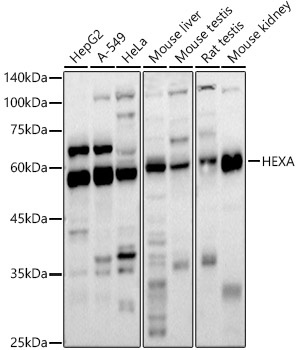
Western blot analysis of various lysates using HEXA Rabbit pAb (CAB5646) at 1:1000 dilution. Secondary antibody: HRP-conjugated Goat anti-Rabbit IgG (H+L) (CABS014) at 1:10000 dilution. Lysates/proteins: 25μg per lane. Blocking buffer: 3% nonfat dry milk in TBST. Detection: ECL Basic Kit (AbGn00020). Exposure time: 30s.
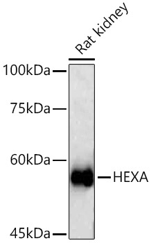
Western blot analysis of lysates from Rat kidney using HEXA Rabbit pAb (CAB5646) at 1:500 dilution incubated overnight at 4℃. Secondary antibody: HRP-conjugated Goat anti-Rabbit IgG (H+L) (CABS014) at 1:10000 dilution. Lysates/proteins: 25 μg per lane. Blocking buffer: 3% nonfat dry milk in TBST. Detection: ECL Basic Kit (AbGn00020). Exposure time: 30s.
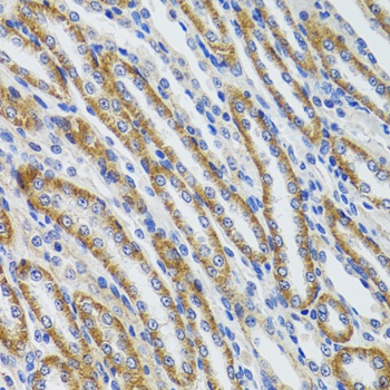
Immunohistochemistry analysis of paraffin-embedded Mouse kidney using HEXA Rabbit pAb (CAB5646) at dilution of 1:100 (40x lens). Microwave antigen retrieval performed with 0.01M PBS Buffer (pH 7.2) prior to IHC staining.
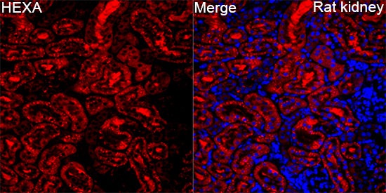
Immunofluorescence analysis of paraffin-embedded Rat kidney tissue using HEXA Rabbit pAb(CAB5646) at a dilution of 1:100 (40x lens). Secondary antibody:Cy3 Goat Anti-Rabbit IgG (H+L)(CABS007) at 1:500 dilution. Blue: DAPI for nuclear staining. Perform high pressure antigen retrieval with 0.01 M citrate buffer (pH 6.0) prior to IF staining.