The HIF3A Antibody (CAB20020) is a high-quality antibody developed for reliable detection and analysis of target proteins. This antibody, produced in rabbits, exhibits high specificity and sensitivity for human samples, making it suitable for Western blot analysis and other immunological techniques.HIF3A is a key regulator of cellular responses to hypoxia, playing a role in various physiological and pathological processes such as angiogenesis, metabolism, and cancer progression. By targeting HIF3A, researchers can gain insights into these processes and potentially develop new strategies for treating hypoxia-related diseases.
This antibody is validated for use in WB, IF/ICC, ELISA applications and has demonstrated reactivity against Human, Mouse, Rat samples.
Product Name:
HIF3A Antibody
SKU:
CAB20020
Size:
20μL, 100μL
Reactivity:
Human, Mouse, Rat
Conjugate:
Unconjugated
Immunogen:
Recombinant protein (or fragment).This information is considered to be commercially sensitive.
The protein encoded by this gene is the alpha-3 subunit of one of several alpha/beta-subunit heterodimeric transcription factors that regulate many adaptive responses to low oxygen tension (hypoxia). The alpha-3 subunit lacks the transactivation domain found in factors containing either the alpha-1 or alpha-2 subunits. It is thought that factors containing the alpha-3 subunit are negative regulators of hypoxia-inducible gene expression. Multiple alternatively spliced transcript variants have been found for this gene.
Purification Method
Affinity purification
Gene ID
64344
RRID
AB_2862923
Buffer Information
Store at -20℃. Avoid freeze / thaw cycles. Buffer: PBS containing 50% glycerol, preserved with proclin300 or sodium azide, pH 7.3.
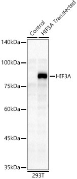
Western blot analysis of lysates from 293F-HIF3A-FlagN cells, using HIF3A Rabbit pAb (CAB20020) at 1:1000 dilution. Secondary antibody: HRP-conjugated Goat anti-Rabbit IgG (H+L) (CABS014) at 1:10000 dilution. Lysates/proteins: 25μg per lane. Blocking buffer: 3% nonfat dry milk in TBST. Detection: ECL Basic Kit (AbGn00020). Exposure time: 30s.
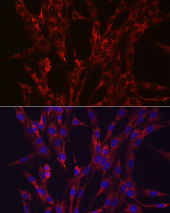
Immunofluorescence analysis of C6 cells using HIF3A Rabbit pAb (CAB20020) at dilution of 1:100 (40x lens). Secondary antibody: Cy3-conjugated Goat anti-Rabbit IgG (H+L) (CABS007) at 1:500 dilution. Blue: DAPI for nuclear staining.