The HIPK2 Monoclonal Antibody (CAB9552) is a high-quality antibody developed for reliable detection and analysis of target proteins. This antibody, produced in rabbits, is highly specific to human samples and is validated for use in various applications such as Western blotting and immunofluorescence.HIPK2 is a critical protein kinase that plays a crucial role in regulating cell growth, survival, and stress responses. Dysregulation of HIPK2 has been implicated in various diseases, including cancer and neurodegenerative disorders.
This antibody is validated for use in WB, IF/ICC, ELISA, IF-P applications and has demonstrated reactivity against Human, Mouse samples.
Product Name:
HIPK2 Monoclonal Antibody
SKU:
CAB9552
Size:
20μL, 100μL
Reactivity:
Human, Mouse
Clone Number:
ARC1631
Conjugate:
Unconjugated
Immunogen:
Synthetic peptide. This information is considered to be commercially sensitive.
Recommended starting concentration is 1 μg/mL. Please optimize the concentration based on your specific assay requirements.
Synonyms:
PRO0593, hHIPk2, HIPK2
Positive Sample:
RD, Raji
Cellular Localization:
Cytoplasm, Nucleus, Pml Body.
Calculated MW:
101KDa/128KDa/131kDa
Observed MW:
128kDa/140kDa
This gene encodes a conserved serine/threonine kinase that is a member of the homeodomain-interacting protein kinase family. The encoded protein interacts with homeodomain transcription factors and many other transcription factors such as p53, and can function as both a corepressor and a coactivator depending on the transcription factor and its subcellular localization. Multiple transcript variants encoding different isoforms have been found for this gene.
Purification Method
Affinity purification
Gene ID
28996
RRID
AB_2863722
Buffer Information
Store at -20℃. Avoid freeze / thaw cycles. Buffer: PBS containing 50% glycerol and 0.05% BSA, preserved with proclin300 or sodium azide, pH 7.3.
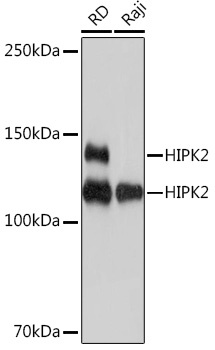
Western blot analysis of various lysates using HIPK2 Rabbit mAb (CAB9552) at 1:1000 dilution. Secondary antibody: HRP-conjugated Goat anti-Rabbit IgG (H+L) (CABS014) at 1:10000 dilution. Lysates/proteins: 25μg per lane. Blocking buffer: 3% nonfat dry milk in TBST. Detection: ECL Basic Kit (AbGn00020). Exposure time: 1s.
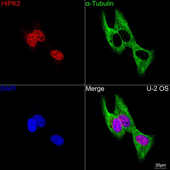
Confocal imaging of U-2 OS cells using HIPK2 Rabbit mAb (CAB9552, dilution 1:200) followed by a further incubation with Cy3 Goat Anti-Rabbit IgG (H+L) (CABS007, dilution 1:500) (Red). The cells were counterstained with α-Tubulin Mouse mAb (AC012, dilution 1:400) followed by incubation with ABflo® 488-conjugated Goat Anti-Mouse IgG (H+L) Ab (CABS076, dilution 1:500) (Green). DAPI was used for nuclear staining (Blue). Objective: 100x.
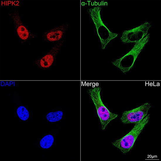
Confocal imaging of HeLa cells using HIPK2 Rabbit mAb (CAB9552, dilution 1:200) followed by a further incubation with Cy3 Goat Anti-Rabbit IgG (H+L) (CABS007, dilution 1:500) (Red). The cells were counterstained with α-Tubulin Mouse mAb (AC012, dilution 1:400) followed by incubation with ABflo® 488-conjugated Goat Anti-Mouse IgG (H+L) Ab (CABS076, dilution 1:500) (Green). DAPI was used for nuclear staining (Blue). Objective: 100x.