The HMGCS1 Antibody (CAB3916) is a high-quality antibody developed for reliable detection and analysis of target proteins. This antibody, raised in rabbits, is highly specific to human samples and is validated for use in Western blot applications. By binding to the HMGCS1 protein, this antibody allows for the accurate detection and analysis of HMGCS1 in various cell types, making it an ideal choice for studies in metabolism, lipid metabolism, and metabolic diseases.HMGCS1 plays a crucial role in energy metabolism, specifically in the production of ketone bodies during times of fasting or low carbohydrate intake.
This antibody is validated for use in WB, IF/ICC, ELISA applications and has demonstrated reactivity against Human, Mouse, Rat samples.
Product Name:
HMGCS1 Antibody
SKU:
CAB3916
Size:
20μL, 100μL
Reactivity:
Human, Mouse, Rat
Conjugate:
Unconjugated
Immunogen:
Recombinant protein (or fragment).This information is considered to be commercially sensitive.
Recommended starting concentration is 1 μg/mL. Please optimize the concentration based on your specific assay requirements.
Synonyms:
HMGCS, HMGCS1
Positive Sample:
NCI-H460, HeLa, Jurkat, Mouse kidney, Mouse liver, Rat liver, Rat spinal cord
Cellular Localization:
Cytoplasm.
Calculated MW:
57kDa
Observed MW:
57kDa
Enables protein homodimerization activity. Predicted to be involved in acetyl-CoA metabolic process and farnesyl diphosphate biosynthetic process, mevalonate pathway. Predicted to be located in cytoplasm. Predicted to be active in cytosol.
Purification Method
Affinity purification
Gene ID
3157
RRID
AB_2765382
Buffer Information
Store at -20℃. Avoid freeze / thaw cycles. Buffer: PBS with 0.01% thimerosal,50% glycerol,pH7.3.
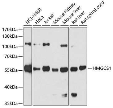
Western blot analysis of various lysates using HMGCS1 Rabbit pAb (CAB3916) at 1:7000 dilution. Secondary antibody: HRP-conjugated Goat anti-Rabbit IgG (H+L) (CABS014) at 1:10000 dilution. Lysates/proteins: 25μg per lane. Blocking buffer: 3% nonfat dry milk in TBST. Detection: ECL Basic Kit (AbGn00020). Exposure time: 30s.
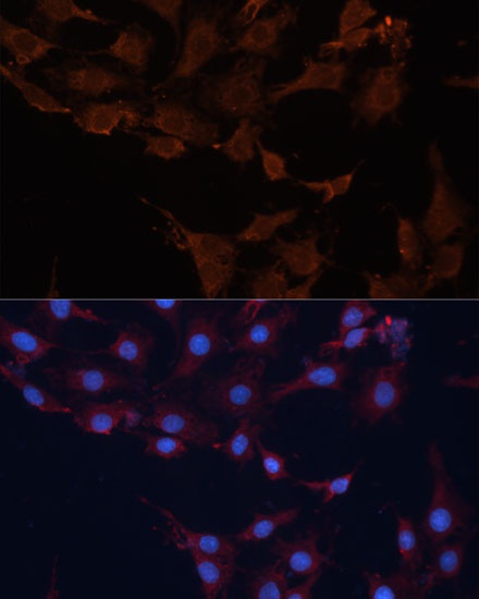
Immunofluorescence analysis of C6 cells using HMGCS1 Rabbit pAb (CAB3916) at dilution of 1:100 (40x lens). Secondary antibody: Cy3-conjugated Goat anti-Rabbit IgG (H+L) (CABS007) at 1:500 dilution. Blue: DAPI for nuclear staining.