The Heme Oxygenase 1 (HO-1/HMOX1) Antibody (CAB11102) is a high-quality antibody developed for reliable detection and analysis of target proteins. Raised in rabbits, this antibody is highly specific for human samples and has been validated for use in Western blot applications. By binding to the HO-1 protein, this antibody allows for accurate detection and analysis in a variety of cell types, making it a valuable asset for investigations in oxidative stress, inflammation, and diseases like cancer and cardiovascular disorders.HO-1, a stress-responsive enzyme, plays a crucial role in the breakdown of heme into biliverdin, carbon monoxide, and iron, leading to the antioxidant and cytoprotective effects that are essential for cellular survival under oxidative conditions.
This antibody is validated for use in WB, IHC-P, ELISA, IF-P applications and has demonstrated reactivity against Human, Mouse, Rat samples.
Product Name:
Heme Oxygenase 1 (HO-1/HMOX1) Antibody
SKU:
CAB11102
Size:
20μL, 100μL
Reactivity:
Human, Mouse, Rat
Conjugate:
Unconjugated
Immunogen:
Recombinant protein (or fragment).This information is considered to be commercially sensitive.
Heme oxygenase, an essential enzyme in heme catabolism, cleaves heme to form biliverdin, which is subsequently converted to bilirubin by biliverdin reductase, and carbon monoxide, a putative neurotransmitter. Heme oxygenase activity is induced by its substrate heme and by various nonheme substances. Heme oxygenase occurs as 2 isozymes, an inducible heme oxygenase-1 and a constitutive heme oxygenase-2. HMOX1 and HMOX2 belong to the heme oxygenase family.
Purification Method
Affinity purification
Gene ID
3162
RRID
AB_2758406
Buffer Information
Store at -20℃. Avoid freeze / thaw cycles. Buffer: PBS with 0.09% Sodium azide,50% glycerol,pH7.3.
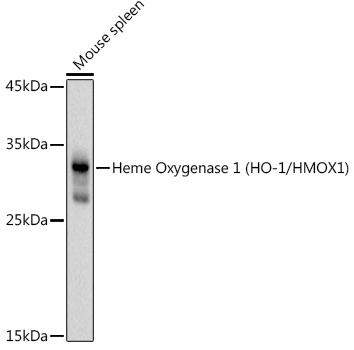
Western blot analysis of lysates from Mouse spleen, using Heme Oxygenase 1 (HO-1/HMOX1) Rabbit pAb (CAB11102) at 1:1000 dilution. Secondary antibody: HRP-conjugated Goat anti-Rabbit IgG (H+L) (CABS014) at 1:10000 dilution. Lysates/proteins: 25μg per lane. Blocking buffer: 3% nonfat dry milk in TBST. Detection: ECL Basic Kit (AbGn00020). Exposure time: 3s.
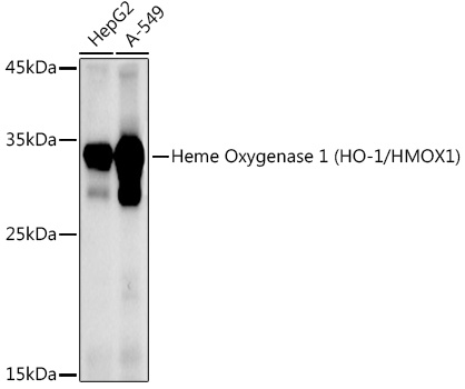
Western blot analysis of various lysates using Heme Oxygenase 1 (HO-1/HMOX1) Rabbit pAb (CAB11102) at 1:1000 dilution. Secondary antibody: HRP-conjugated Goat anti-Rabbit IgG (H+L) (CABS014) at 1:10000 dilution. Lysates/proteins: 25μg per lane. Blocking buffer: 3% nonfat dry milk in TBST. Detection: ECL Enhanced Kit (AbGn00021). Exposure time: 90s.
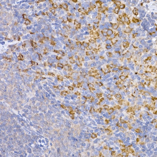
Immunohistochemistry analysis of paraffin-embedded Rat spleen using Heme Oxygenase 1 (HO-1/HMOX1) Rabbit pAb (CAB11102) at dilution of 1:50 (40x lens). High pressure antigen retrieval performed with 0.01M Citrate buffer (pH 6.0) prior to IHC staining.
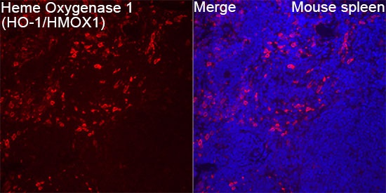
Confocal imaging of paraffin-embedded Mouse spleen tissue using Heme Oxygenase 1 (HO-1/HMOX1) Rabbit pAb (CAB11102, dilution 1:100) followed by a further incubation with Cy3 Goat Anti-Rabbit IgG (H+L) (CABS007, dilution 1:500) (Red). DAPI was used for nuclear staining (Blue). High pressure antigen retrieval performed with 0.01M Citrate Buffer (pH 6.0) prior to IF staining. Objective: 40x.


 ELISA Kit (RTEB0355)")