The Hmox1 Polyclonal Antibody (CAB23650) is a high-quality antibody developed for reliable detection and analysis of target proteins. This antibody, produced using rabbits, demonstrates a high level of reactivity with human samples and has been specifically validated for use in Western blot applications.HMOX1 plays a crucial role in various physiological processes, including antioxidative defense, anti-inflammatory responses, and cell proliferation. Dysregulation of HMOX1 has been linked to numerous pathological conditions, such as cancer, neurodegenerative diseases, and cardiovascular disorders.
This antibody is validated for use in WB, IHC-P, ELISA, IF-P applications and has demonstrated reactivity against Mouse, Rat samples.
Product Name:
Hmox1 Polyclonal Antibody
SKU:
CAB23650
Size:
20μL, 100μL
Reactivity:
Mouse, Rat
Conjugate:
Unconjugated
Immunogen:
Recombinant protein (or fragment).This information is considered to be commercially sensitive.
Recommended starting concentration is 1 μg/mL. Please optimize the concentration based on your specific assay requirements.
Synonyms:
HO1, HO-1, Hmox, Hemox, Hsp32, D8Wsu38e, Hmox1
Positive Sample:
NIH/3T3 treated with arsenite, Mouse spleen, Rat spleen
Cellular Localization:
Endoplasmic Reticulum Membrane.
Calculated MW:
33KDa
Observed MW:
30kDa/35kDa
Predicted to enable several functions, including heme binding activity; heme oxygenase (decyclizing) activity; and protein homodimerization activity. Acts upstream of or within several processes, including cellular response to cisplatin; cellular response to metal ion; and regulation of macroautophagy. Located in nucleus. Is expressed in several structures, including alimentary system; central nervous system; early conceptus; genitourinary system; and sensory organ. Used to study hemochromatosis and malaria. Human ortholog(s) of this gene implicated in several diseases, including artery disease (multiple); cerebrovascular disease (multiple); factor VIII deficiency; lung disease (multiple); and sickle cell anemia. Orthologous to human HMOX1 (heme oxygenase 1).
Purification Method
Affinity purification
Gene ID
15368
Buffer Information
Store at -20℃. Avoid freeze / thaw cycles. Buffer: PBS containing 50% glycerol, preserved with proclin300 or sodium azide, pH 7.3.
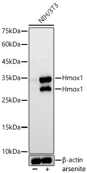
Western blot analysis of lysates from NIH/3T3 cells using Hmox1 Rabbit pAb (CAB23650) at 1:1000 dilution. NIH/3T3 cells were treated with arsenite(50 μM) at 37℃ for 8 hours. Secondary antibody: HRP-conjugated Goat anti-Rabbit IgG (H+L) (CABS014) at 1:10000 dilution. Lysates/proteins: 25 μg per lane. Blocking buffer: 3% nonfat dry milk in TBST. Detection: ECL Basic Kit (AbGn00020). Exposure time: 1s.
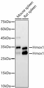
Western blot analysis of various lysates using Hmox1 Rabbit pAb (CAB23650) at 1:1000 dilution. Secondary antibody: HRP-conjugated Goat anti-Rabbit IgG (H+L) (CABS014) at 1:10000 dilution. Lysates / proteins: 25 μg per lane. Blocking buffer: 3 % nonfat dry milk in TBST. Detection: ECL Basic Kit (AbGn00020). Exposure time: 30s.
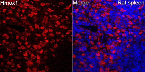
Immunofluorescence analysis of Rat spleen tissue using Hmox1 Rabbit pAb (CAB23650) at a dilution of 1:100 (40x lens). Secondary antibody: Cy3-conjugated Goat anti-Rabbit IgG (H+L)(CABS007) at 1:500 dilution. Blue: DAPI for nuclear staining. High pressure antigen retrieval performed with 0.01M Citrate Buffer(pH 6.0) prior to IF staining.
")
")


