The hnRNP Q/SYNCRIP Monoclonal Antibody (CAB9609) is a high-quality antibody developed for reliable detection and analysis of target proteins. This antibody, produced in rabbits, is highly specific and reactive with human samples, making it a valuable tool for studying HNRNP Q in different cell types.Validated for use in techniques such as Western blotting, the HNRNP Q Rabbit Monoclonal Antibody (CAB9609) binds specifically to the HNRNP Q protein, allowing for accurate detection and analysis. Its application in immunology and cancer research is particularly significant, as HNRNP Q has been implicated in processes such as alternative splicing in cancer cells and regulation of gene expression in immune responses.
This antibody is validated for use in WB, IHC-P, IF/ICC, ELISA applications and has demonstrated reactivity against Human, Mouse, Rat samples.
Product Name:
hnRNP Q/SYNCRIP Monoclonal Antibody
SKU:
CAB9609
Size:
20μL, 100μL
Reactivity:
Human, Mouse, Rat
Clone Number:
ARC1656
Conjugate:
Unconjugated
Immunogen:
Synthetic peptide. This information is considered to be commercially sensitive.
This gene encodes a member of the cellular heterogeneous nuclear ribonucleoprotein (hnRNP) family. hnRNPs are RNA binding proteins that complex with heterogeneous nuclear RNA (hnRNA) and regulate alternative splicing, polyadenylation, and other aspects of mRNA metabolism and transport. The encoded protein plays a role in multiple aspects of mRNA maturation and is associated with several multiprotein complexes including the apoB RNA editing-complex and survival of motor neurons (SMN) complex. Alternatively spliced transcript variants encoding multiple isoforms have been observed for this gene, and a pseudogene of this gene is located on the short arm of chromosome 20.
Purification Method
Affinity purification
Gene ID
10492
RRID
AB_2863736
Buffer Information
Store at -20℃. Avoid freeze / thaw cycles. Buffer: PBS containing 50% glycerol and 0.05% BSA, preserved with proclin300 or sodium azide, pH 7.3.
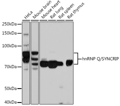
Western blot analysis of various lysates using hnRNP Q/SYNCRIP /SYNCRIP Rabbit mAb (CAB9609) at 1:1000 dilution. Secondary antibody: HRP-conjugated Goat anti-Rabbit IgG (H+L) (CABS014) at 1:10000 dilution. Lysates/proteins: 25μg per lane. Blocking buffer: 3% nonfat dry milk in TBST. Detection: ECL Basic Kit (AbGn00020). Exposure time: 3s.
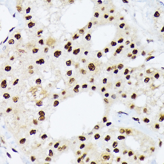
Immunohistochemistry analysis of paraffin-embedded Human liver cancer using hnRNP Q/SYNCRIP /SYNCRIP Rabbit mAb (CAB9609) at dilution of 1:100 (40x lens). Microwave antigen retrieval performed with 0.01M Tris/EDTA Buffer (pH 9.0) prior to IHC staining.
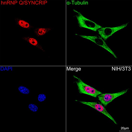
Confocal imaging of NIH/3T3 cells using hnRNP Q/SYNCRIP Rabbit mAb (CAB9609, dilution 1:200) followed by a further incubation with Cy3 Goat Anti-Rabbit IgG (H+L) (CABS007, dilution 1:500) (Red). The cells were counterstained with α-Tubulin Mouse mAb (AC012, dilution 1:400) followed by incubation with ABflo® 488-conjugated Goat Anti-Mouse IgG (H+L) Ab (CABS076, dilution 1:500) (Green). DAPI was used for nuclear staining (Blue). Objective: 100x.
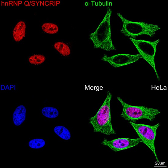
Confocal imaging of HeLa cells using hnRNP Q/SYNCRIP Rabbit mAb (CAB9609, dilution 1:200) followed by a further incubation with Cy3 Goat Anti-Rabbit IgG (H+L) (CABS007, dilution 1:500) (Red). The cells were counterstained with α-Tubulin Mouse mAb (AC012, dilution 1:400) followed by incubation with ABflo® 488-conjugated Goat Anti-Mouse IgG (H+L) Ab (CABS076, dilution 1:500) (Green). DAPI was used for nuclear staining (Blue). Objective: 100x.