The HNRNPH1 Antibody (CAB5924) is a high-quality antibody developed for reliable detection and analysis of target proteins. This antibody, generated in rabbits, has high specificity for human samples and has been validated for use in Western blot applications.HNRNPH1 plays a crucial role in the regulation of gene expression and alternative splicing, making it a key player in various biological processes. Its dysregulation has been linked to diseases such as cancer, neurodevelopmental disorders, and autoimmune conditions, highlighting the importance of studying its function and mechanism of action.
This antibody is validated for use in WB, IHC-P, IF/ICC, ELISA applications and has demonstrated reactivity against Human, Mouse, Rat samples.
Product Name:
HNRNPH1 Antibody
SKU:
CAB5924
Size:
20μL, 100μL
Reactivity:
Human, Mouse, Rat
Conjugate:
Unconjugated
Immunogen:
Recombinant protein (or fragment).This information is considered to be commercially sensitive.
Recommended starting concentration is 1 μg/mL. Please optimize the concentration based on your specific assay requirements.
Synonyms:
HNRPH, HNRPH1, NEDCDS, hnRNPH, HNRNPH1
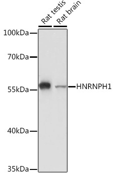
Positive Sample:
Rat testis, Rat brain
Cellular Localization:
Nucleus, Nucleoplasm.
Calculated MW:
49kDa
Observed MW:
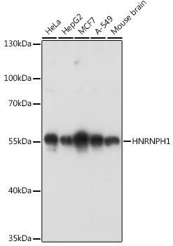
55kDa
This gene encodes a member of a subfamily of ubiquitously expressed heterogeneous nuclear ribonucleoproteins (hnRNPs). The hnRNPs are RNA binding proteins that complex with heterogeneous nuclear RNA. These proteins are associated with pre-mRNAs in the nucleus and appear to influence pre-mRNA processing and other aspects of mRNA metabolism and transport. While all of the hnRNPs are present in the nucleus, some may shuttle between the nucleus and the cytoplasm. The hnRNP proteins have distinct nucleic acid binding properties. The protein encoded by this gene has three repeats of quasi-RRM domains that bind to RNA and is very similar to the family member HNRPF. This gene may be associated with hereditary lymphedema type I. Alternatively spliced transcript variants have been described
Purification Method
Affinity purification
Gene ID
3187
RRID
AB_2766663
Buffer Information
Store at -20℃. Avoid freeze / thaw cycles. Buffer: PBS with 0.01% thimerosal,50% glycerol,pH7.3.
Western blot analysis of various lysates using HNRNPH1 Rabbit pAb (CAB5924) at 1:3000 dilution. Secondary antibody: HRP-conjugated Goat anti-Rabbit IgG (H+L) (CABS014) at 1:10000 dilution. Lysates/proteins: 25μg per lane. Blocking buffer: 3% nonfat dry milk in TBST. Detection: ECL Basic Kit (AbGn00020). Exposure time: 90s.
Western blot analysis of various lysates using HNRNPH1 Rabbit pAb (CAB5924) at 1:3000 dilution. Secondary antibody: HRP-conjugated Goat anti-Rabbit IgG (H+L) (CABS014) at 1:10000 dilution. Lysates/proteins: 25μg per lane. Blocking buffer: 3% nonfat dry milk in TBST. Detection: ECL Basic Kit (AbGn00020). Exposure time: 90s.
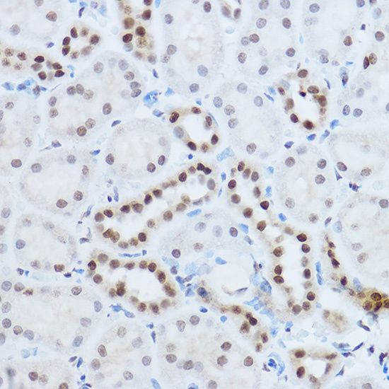
Immunohistochemistry analysis of paraffin-embedded Mouse kidney using HNRNPH1 Rabbit pAb (CAB5924) at dilution of 1:100 (40x lens). High pressure antigen retrieval performed with 0.01M Citrate buffer (pH 6.0) prior to IHC staining.
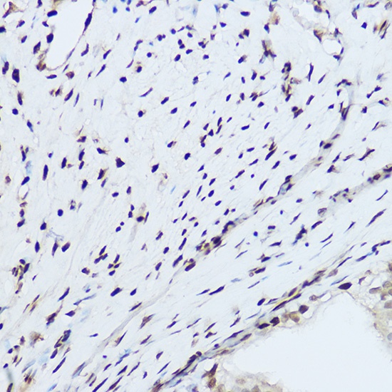
Immunohistochemistry analysis of paraffin-embedded Rat ovary using HNRNPH1 Rabbit pAb (CAB5924) at dilution of 1:100 (40x lens). High pressure antigen retrieval performed with 0.01M Citrate buffer (pH 6.0) prior to IHC staining.
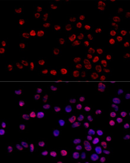
Immunofluorescence analysis of 293T cells using HNRNPH1 Rabbit pAb (CAB5924) at dilution of 1:100 (40x lens). Secondary antibody: Cy3-conjugated Goat anti-Rabbit IgG (H+L) (CABS007) at 1:500 dilution. Blue: DAPI for nuclear staining.