The HOOK3 Antibody (CAB15536) is a high-quality antibody developed for reliable detection and analysis of target proteins. This antibody, generated in rabbits, exhibits high specificity and sensitivity towards human samples, making it ideal for Western blot analysis.HOOK3 is involved in various cellular processes, including intracellular transport, ciliary biogenesis, and cell division. Its dysfunction has been linked to developmental disorders and neurodegenerative diseases, making it a crucial target for investigation in the fields of cell biology and neuroscience.
This antibody is validated for use in WB, IF/ICC, ELISA applications and has demonstrated reactivity against Human, Mouse, Rat samples.
Product Name:
HOOK3 Antibody
SKU:
CAB15536
Size:
20μL, 100μL
Reactivity:
Human, Mouse, Rat
Conjugate:
Unconjugated
Immunogen:
Recombinant protein (or fragment).This information is considered to be commercially sensitive.
Recommended starting concentration is 1 μg/mL. Please optimize the concentration based on your specific assay requirements.
Synonyms:
HK3, HOOK3
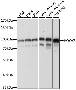
Positive Sample:
LO2, HeLa, 293T, mouse heart, mouse kidney, rat lung
Cellular Localization:
Cytoplasm, Golgi Apparatus, Cytoskeleton.
Calculated MW:
83kDa
Observed MW:
170kDa
Hook proteins are cytosolic coiled-coil proteins that contain conserved N-terminal domains, which attach to microtubules, and more divergent C-terminal domains, which mediate binding to organelles. The Drosophila Hook protein is a component of the endocytic compartment.
Purification Method
Affinity purification
Gene ID
84376
RRID
AB_2762937
Buffer Information
Store at -20℃. Avoid freeze / thaw cycles. Buffer: PBS with 0.01% thimerosal,50% glycerol,pH7.3.
Western blot analysis of various lysates using HOOK3 Rabbit pAb (CAB15536) at 1:1000 dilution. Secondary antibody: HRP-conjugated Goat anti-Rabbit IgG (H+L) (CABS014) at 1:10000 dilution. Lysates/proteins: 25μg per lane. Blocking buffer: 3% nonfat dry milk in TBST. Detection: ECL Basic Kit (AbGn00020). Exposure time: 60s.
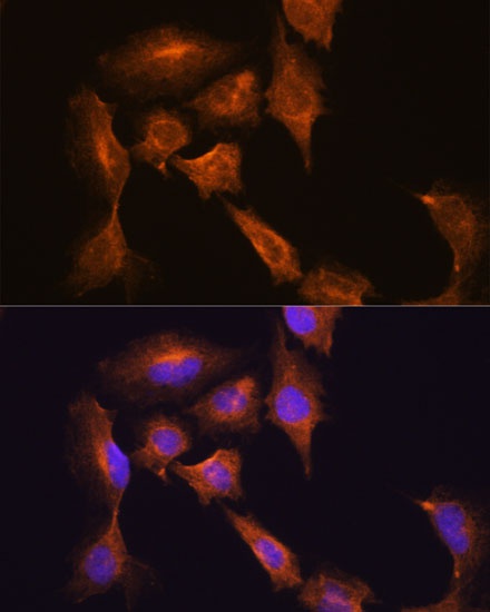
Immunofluorescence analysis of C6 cells using HOOK3 Rabbit pAb (CAB15536) at dilution of 1:100. Secondary antibody: Cy3-conjugated Goat anti-Rabbit IgG (H+L) (CABS007) at 1:500 dilution. Blue: DAPI for nuclear staining.