The HOXB1 Antibody (CAB6619) is a high-quality antibody developed for reliable detection and analysis of target proteins. This antibody, produced in rabbits, shows high reactivity with human samples and has been validated for use in Western blot applications. By binding specifically to the HOXB1 protein, this antibody allows for the precise detection and analysis of HOXB1 expression in a variety of cell types.HOXB1 is a crucial regulator of gene expression during early development, playing a key role in specifying the identity of cells in the developing embryo.
This antibody is validated for use in WB, IF/ICC, ELISA applications and has demonstrated reactivity against Human, Mouse, Rat samples.
Product Name:
HOXB1 Antibody
SKU:
CAB6619
Size:
20μL, 100μL
Reactivity:
Human, Mouse, Rat
Conjugate:
Unconjugated
Immunogen:
Recombinant protein (or fragment).This information is considered to be commercially sensitive.
Recommended starting concentration is 1 μg/mL. Please optimize the concentration based on your specific assay requirements.
Synonyms:
HOX2, HCFP3, HOX2I, Hox-2.9, HOXB1
Positive Sample:
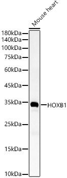
Mouse heart
Cellular Localization:
Nucleus.
Calculated MW:
32kDa
Observed MW:
32kDa
This gene belongs to the homeobox family of genes. The homeobox genes encode a highly conserved family of transcription factors that play an important role in morphogenesis in all multicellular organisms. Mammals possess four similar homeobox gene clusters, HOXA, HOXB, HOXC and HOXD, located on different chromosomes, consisting of 9 to 11 genes arranged in tandem. This gene is one of several homeobox HOXB genes located in a cluster on chromosome 17.
Purification Method
Affinity purification
Gene ID
3211
RRID
AB_2767209
Buffer Information
Store at -20℃. Avoid freeze / thaw cycles. Buffer: PBS containing 50% glycerol, preserved with proclin300 or sodium azide, pH 7.3.
Western blot analysis of lysates from Mouse heart, using HOXB1 Rabbit pAb (CAB6619) at 1:4000 dilution. Secondary antibody: HRP-conjugated Goat anti-Rabbit IgG (H+L) (CABS014) at 1:10000 dilution. Lysates/proteins: 25μg per lane. Blocking buffer: 3% nonfat dry milk in TBST. Detection: ECL Basic Kit (AbGn00020). Exposure time: 30s.
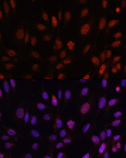
Immunofluorescence analysis of U-2 OS cells using HOXB1 Rabbit pAb (CAB6619) at dilution of 1:100. Secondary antibody: Cy3-conjugated Goat anti-Rabbit IgG (H+L) (CABS007) at 1:500 dilution. Blue: DAPI for nuclear staining.