The HPD Antibody (CAB6505) is a high-quality antibody developed for reliable detection and analysis of target proteins. This antibody, raised in rabbits, is highly specific to human samples and has been validated for use in Western blot applications.The HPD protein, also known as 4-hydroxyphenylpyruvate dioxygenase, plays a crucial role in the tyrosine catabolic pathway and is essential for the breakdown of tyrosine into various metabolites.
This antibody is validated for use in WB, IHC-P, ELISA applications and has demonstrated reactivity against Human, Mouse, Rat samples.
Product Name:
HPD Antibody
SKU:
CAB6505
Size:
20μL, 100μL
Reactivity:
Human, Mouse, Rat
Conjugate:
Unconjugated
Immunogen:
Recombinant protein (or fragment).This information is considered to be commercially sensitive.
Recommended starting concentration is 1 μg/mL. Please optimize the concentration based on your specific assay requirements.
Synonyms:
PPD, 4HPPD, GLOD3, 4-HPPD, HPPDASE, HPD
Positive Sample:
HL-60, Mouse liver, Mouse kidney, Rat liver, Rat kidney
Cellular Localization:
Cytosol, Extracellular Exosome.
Calculated MW:
45kDa
Observed MW:
45kDa
The protein encoded by this gene is an enzyme in the catabolic pathway of tyrosine. The encoded protein catalyzes the conversion of 4-hydroxyphenylpyruvate to homogentisate. Defects in this gene are a cause of tyrosinemia type 3 (TYRO3) and hawkinsinuria (HAWK). Two transcript variants encoding different isoforms have been found for this gene.
Purification Method
Affinity purification
Gene ID
3242
RRID
AB_2767101
Buffer Information
Store at -20℃. Avoid freeze / thaw cycles. Buffer: PBS containing 50% glycerol, preserved with proclin300 or sodium azide, pH 7.3.
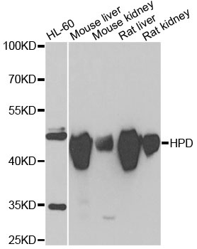
Western blot analysis of various lysates using HPD Rabbit pAb (CAB6505) at 1:1000 dilution. Secondary antibody: HRP-conjugated Goat anti-Rabbit IgG (H+L) (CABS014) at 1:10000 dilution. Lysates/proteins: 25μg per lane. Blocking buffer: 3% nonfat dry milk in TBST. Detection: ECL Enhanced Kit (AbGn00021). Exposure time: 10s.
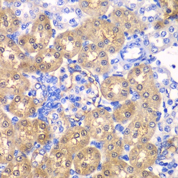
Immunohistochemistry analysis of paraffin-embedded Rat kidney using HPD Rabbit pAb (CAB6505) at dilution of 1:100 (40x lens). Microwave antigen retrieval performed with 0.01M PBS Buffer (pH 7.2) prior to IHC staining.