The HSP27/HSPB1 Monoclonal Antibody (CAB11156) is a high-quality antibody developed for reliable detection and analysis of target proteins. This highly reactive antibody is specifically designed for use in Western blot applications and is validated for detection of the HSP27 protein in human samples.HSP27 is known to play a role in regulating cell survival, apoptosis, and cellular protection under stressful conditions. Its involvement in various cellular processes makes it a key target for research in areas such as cancer, neurodegenerative diseases, and cardiovascular disorders.
This antibody is validated for use in WB, IF/ICC, ELISA, IF-P applications and has demonstrated reactivity against Human, Mouse, Rat samples.
Product Name:
HSP27/HSPB1 Monoclonal Antibody
SKU:
CAB11156
Size:
20μL, 100μL
Reactivity:
Human, Mouse, Rat
Clone Number:
ARC0531
Conjugate:
Unconjugated
Immunogen:
Synthetic peptide. This information is considered to be commercially sensitive.
This gene encodes a member of the small heat shock protein (HSP20) family of proteins. In response to environmental stress, the encoded protein translocates from the cytoplasm to the nucleus and functions as a molecular chaperone that promotes the correct folding of other proteins. This protein plays an important role in the differentiation of a wide variety of cell types. Expression of this gene is correlated with poor clinical outcome in multiple human cancers, and the encoded protein may promote cancer cell proliferation and metastasis, while protecting cancer cells from apoptosis. Mutations in this gene have been identified in human patients with Charcot-Marie-Tooth disease and distal hereditary motor neuropathy.
Purification Method
Affinity purification
Gene ID
3315
RRID
AB_2861510
Buffer Information
Store at -20℃. Avoid freeze / thaw cycles. Buffer: PBS containing 50% glycerol and 0.05% BSA, preserved with proclin300 or sodium azide, pH 7.3.
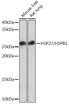
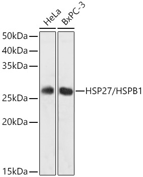
Western blot analysis of various lysates using HSP27/HSPB1Rabbit mAb (CAB11156) at 1:1000 dilution. Secondary antibody: HRP-conjugated Goat anti-Rabbit IgG (H+L) (CABS014) at 1:10000 dilution. Lysates/proteins: 25μg per lane. Blocking buffer: 3% nonfat dry milk in TBST. Detection: ECL Basic Kit (AbGn00020). Exposure time: 30s.
Western blot analysis of various lysates using HSP27/HSPB1Rabbit mAb (CAB11156) at 1:1000 dilution. Secondary antibody: HRP-conjugated Goat anti-Rabbit IgG (H+L) (CABS014) at 1:10000 dilution. Lysates/proteins: 25μg per lane. Blocking buffer: 3% nonfat dry milk in TBST. Detection: ECL Basic Kit (AbGn00020). Exposure time: 1s.
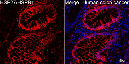
Confocal imaging of paraffin-embedded Human colon cancer tissue using HSP27/HSPB1 Rabbit mAb (CAB11156, dilution 1:200) followed by a further incubation with Cy3 Goat Anti-Rabbit IgG (H+L) (CABS007, dilution 1:500) (Red). DAPI was used for nuclear staining (Blue). Objective: 40x. Perform high pressure antigen retrieval with 0.01 M citrate buffer (pH 6.0) prior to IF staining.
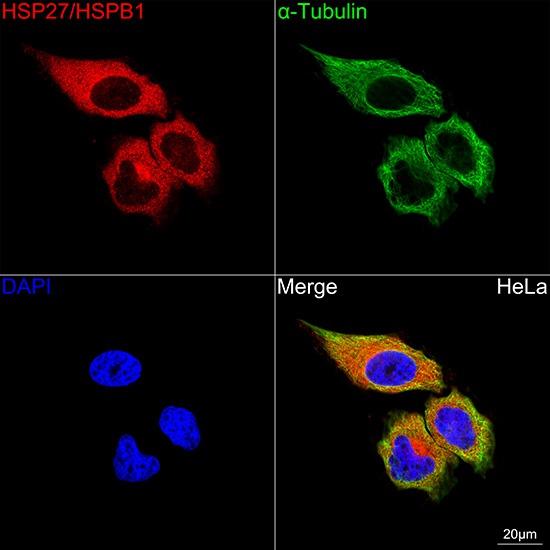
Confocal imaging of HeLa cells using HSP27/HSPB1 Rabbit mAb (CAB11156, dilution 1:200) followed by a further incubation with Cy3 Goat Anti-Rabbit IgG (H+L) (CABS007, dilution 1:500) (Red). The cells were counterstained with α-Tubulin Mouse mAb (AC012, dilution 1:400) followed by incubation with ABflo® 488-conjugated Goat Anti-Mouse IgG (H+L) Ab (CABS076, dilution 1:500) (Green). DAPI was used for nuclear staining (Blue). Objective: 100x.