The HSP60/HSPD1 Monoclonal Antibody (CAB0564) is a high-quality antibody developed for reliable detection and analysis of target proteins. This gene encodes a member of the chaperonin family. The encoded mitochondrial protein may function as a signaling molecule in the innate immune system. This protein is essential for the folding and assembly of newly imported proteins in the mitochondria. This gene is adjacent to a related family member and the region between the 2 genes functions as a bidirectional promoter. Several pseudogenes have been associated with this gene. Two transcript variants encoding the same protein have been identified for this gene. Mutations associated with this gene cause autosomal recessive spastic paraplegia 13.
This antibody is validated for use in WB, IHC-P, IF/ICC, ELISA applications and has demonstrated reactivity against Human, Mouse, Rat, Wheat samples.
Product Name:
HSP60/HSPD1 Monoclonal Antibody
SKU:
CAB0564
Size:
100μL, 20μL
Reactivity:
Human, Mouse, Rat, Wheat
Clone Number:
ARC0260
Conjugate:
Unconjugated
Immunogen:
A synthetic peptide corresponding to a sequence within amino acids 350-450 of human HSP60/HSPD1 (P10809).
Tested Applications:
WBIHC-PIF/ICCELISA
Recommended Dilution:
WB
1:5000 - 1:30000
IHC-P
1:2000 - 1:8000
IF/ICC
1:100 - 1:1000
ELISA
Recommended starting concentration is 1 μg/mL. Please optimize the concentration based on your specific assay requirements.
This gene encodes a member of the chaperonin family. The encoded mitochondrial protein may function as a signaling molecule in the innate immune system. This protein is essential for the folding and assembly of newly imported proteins in the mitochondria. This gene is adjacent to a related family member and the region between the 2 genes functions as a bidirectional promoter. Several pseudogenes have been associated with this gene. Two transcript variants encoding the same protein have been identified for this gene. Mutations associated with this gene cause autosomal recessive spastic paraplegia 13.
Purification Method
Affinity purification
Gene ID
3329
RRID
AB_2861465
Buffer Information
Store at -20℃. Avoid freeze / thaw cycles. Buffer: PBS containing 50% glycerol and 0.05% BSA, preserved with proclin300 or sodium azide, pH 7.3.
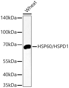
Western blot analysis of lysates from Wheat using HSP60/HSPD1 Rabbit mAb (CAB0564) at 1:10000 dilution incubated overnight at 4℃. Secondary antibody: HRP-conjugated Goat anti-Rabbit IgG (H+L) (AS014) at 1:10000 dilution. Lysates/proteins: 25 μg per lane. Blocking buffer: 3% nonfat dry milk in TBST. Detection: ECL Basic Kit (AbGn00020). Exposure time: 90s.
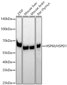
Western blot analysis of various lysates using HSP60/HSPD1 Rabbit mAb (CAB0564) at 1:10000 dilution incubated at room temperature for 1.5 hours. Secondary antibody: HRP-conjugated Goat anti-Rabbit IgG (H+L) (AS014) at 1:10000 dilution. Lysates/proteins: 25 μg per lane. Blocking buffer: 3% nonfat dry milk in TBST. Detection: ECL Basic Kit (AbGn00020). Exposure time: 90s.
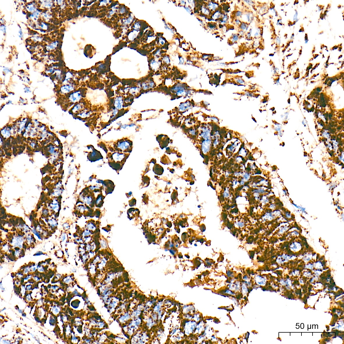
Immunohistochemistry analysis of paraffin-embedded Human colon carcinoma tissue using HSP60/HSPD1 Rabbit mAb (CAB0564) at a dilution of 1:2000 (40x lens). High pressure antigen retrieval performed with 0.01M Tris-EDTA Buffer (pH 9.0) prior to IHC staining.
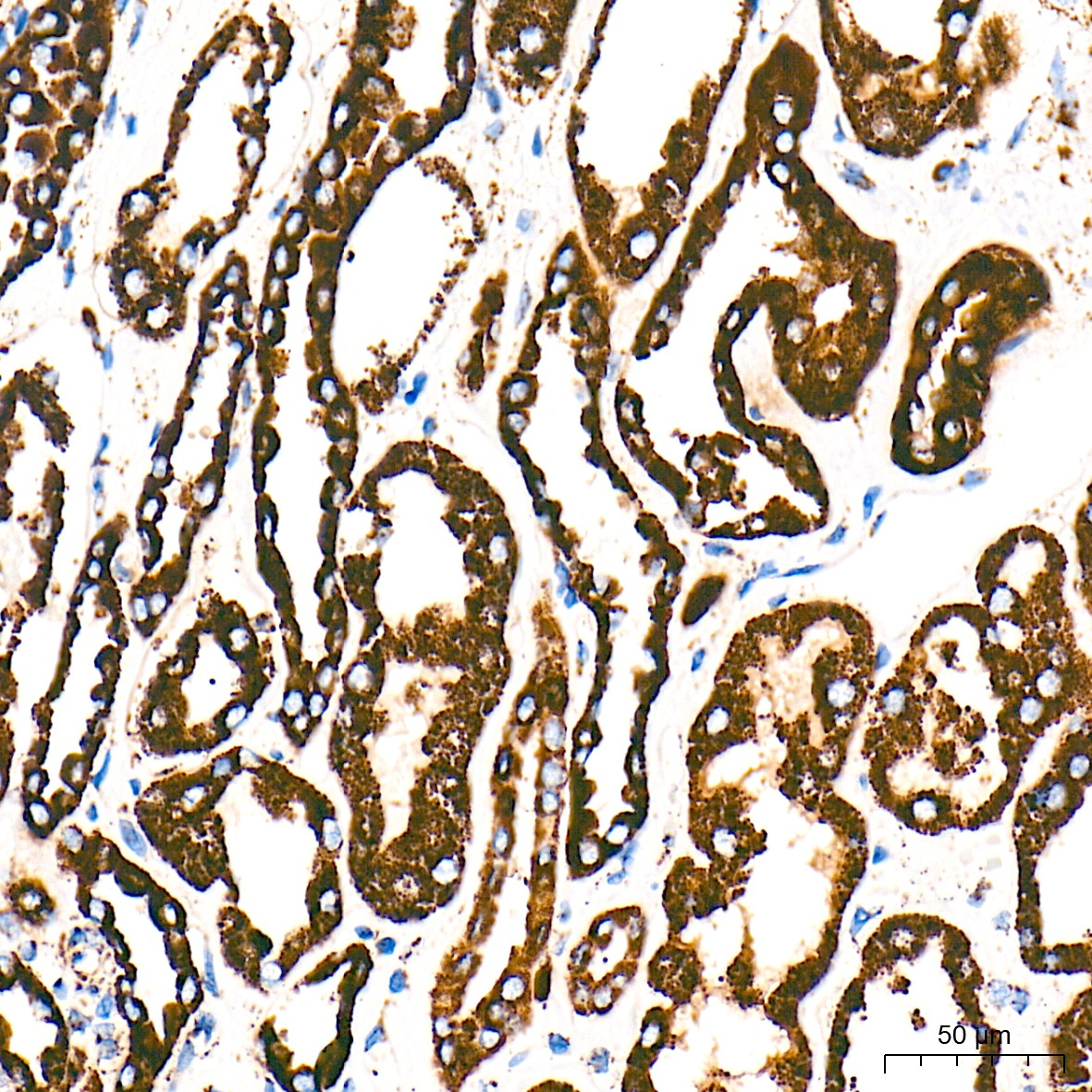
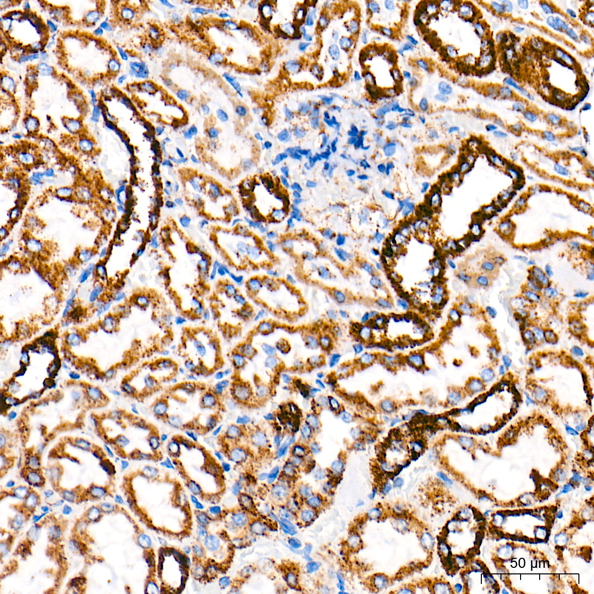
Immunohistochemistry analysis of paraffin-embedded Human kidney tissue using HSP60/HSPD1 Rabbit mAb (CAB0564) at a dilution of 1:2000 (40x lens). High pressure antigen retrieval performed with 0.01M Tris-EDTA Buffer (pH 9.0) prior to IHC staining.
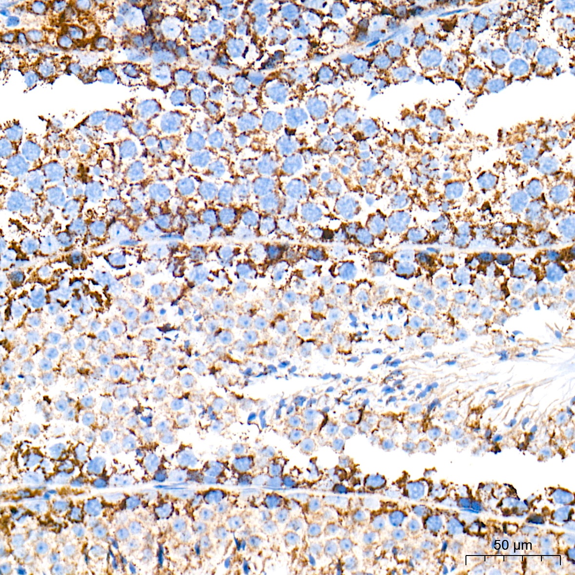
Immunohistochemistry analysis of paraffin-embedded Mouse testis tissue using HSP60/HSPD1 Rabbit mAb (CAB0564) at a dilution of 1:2000 (40x lens). High pressure antigen retrieval performed with 0.01M Tris-EDTA Buffer (pH 9.0) prior to IHC staining.
Immunohistochemistry analysis of paraffin-embedded Rat kidney tissue using HSP60/HSPD1 Rabbit mAb (CAB0564) at a dilution of 1:2000 (40x lens). High pressure antigen retrieval performed with 0.01M Tris-EDTA Buffer (pH 9.0) prior to IHC staining.
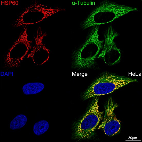
Confocal imaging of HeLa cells using HSP60/HSPD1 Rabbit mAb (CAB0564, dilution 1:100) (Red). The cells were counterstained with α-Tubulin Rabbit mAb (AC049, dilution 1:100) (Green). DAPI was used for nuclear staining (blue). Objective: 60x.
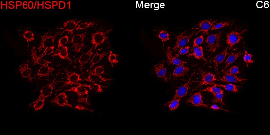
Immunofluorescence analysis of C6 cells using HSP60/HSPD1 Rabbit mAb (CAB0564) at a dilution of 1:100 (40x lens). Secondary antibody: Cy3-conjugated Goat anti-Rabbit IgG (H+L)(AS007) at 1:500 dilution. Blue: DAPI for nuclear staining.


 (RPES0894)")
 (RPES1972)")

