The HTATSF1 Antibody (CAB5977) is a high-quality antibody developed for reliable detection and analysis of target proteins. The protein encoded by this gene functions as a cofactor for the stimulation of transcriptional elongation by HIV-1 Tat, which binds to the HIV-1 promoter through Tat-TAR interaction. This protein may also serve as a dual-function factor to couple transcription and splicing and to facilitate their reciprocal activation. Alternatively spliced transcript variants have been found for this gene.
This antibody is validated for use in WB, IHC-P, IF/ICC, ELISA applications and has demonstrated reactivity against Human, Mouse, Rat samples.
Product Name:
HTATSF1 Antibody
SKU:
CAB5977
Size:
100μL, 20μL
Reactivity:
Human, Mouse, Rat
Conjugate:
Unconjugated
Immunogen:
Recombinant protein (or fragment).This information is considered to be commercially sensitive.
Tested Applications:
WBIHC-PIF/ICCELISA
Recommended Dilution:
WB
1:1000 - 1:2000
IHC-P
1:50 - 1:100
IF/ICC
1:50 - 1:200
ELISA
Recommended starting concentration is 1 μg/mL. Please optimize the concentration based on your specific assay requirements.
Synonyms:
TATSF1, TAT-SF1, dJ196E23.2, HTATSF1
Positive Sample:
U-251MG, Jurkat, HepG2
Cellular Localization:
Nucleus.
Calculated MW:
86kDa
Observed MW:
129kDa
The protein encoded by this gene functions as a cofactor for the stimulation of transcriptional elongation by HIV-1 Tat, which binds to the HIV-1 promoter through Tat-TAR interaction. This protein may also serve as a dual-function factor to couple transcription and splicing and to facilitate their reciprocal activation. Alternatively spliced transcript variants have been found for this gene.
Purification Method
Affinity purification
Gene ID
27336
RRID
AB_2766696
Buffer Information
Store at -20℃. Avoid freeze / thaw cycles. Buffer: PBS containing 50% glycerol, preserved with proclin300 or sodium azide, pH 7.3.
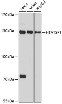
Western blot analysis of various lysates using HTATSF1 Rabbit pAb (CAB5977) at 1:1000 dilution. Secondary antibody: HRP-conjugated Goat anti-Rabbit IgG (H+L) (AS014) at 1:10000 dilution. Lysates/proteins: 25μg per lane. Blocking buffer: 3% nonfat dry milk in TBST. Detection: ECL Basic Kit (AbGn00020). Exposure time: 90s.
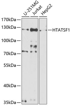
Western blot analysis of various lysates using HTATSF1 Rabbit pAb (CAB5977) at 1:1000 dilution. Secondary antibody: HRP-conjugated Goat anti-Rabbit IgG (H+L) (AS014) at 1:10000 dilution. Lysates/proteins: 25μg per lane. Blocking buffer: 3% nonfat dry milk in TBST. Detection: ECL Basic Kit (AbGn00020). Exposure time: 90s.
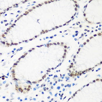
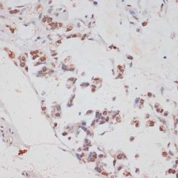
Immunohistochemistry analysis of paraffin-embedded Human stomach using HTATSF1 Rabbit pAb (CAB5977) at dilution of 1:100 (40x lens). Microwave antigen retrieval performed with 0.01M PBS Buffer (pH 7.2) prior to IHC staining.
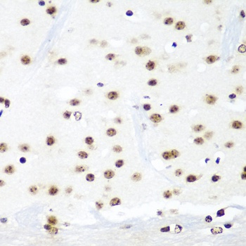
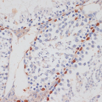
Immunohistochemistry analysis of paraffin-embedded Mouse brain using HTATSF1 Rabbit pAb (CAB5977) at dilution of 1:100 (40x lens). Microwave antigen retrieval performed with 0.01M PBS Buffer (pH 7.2) prior to IHC staining.
Immunohistochemistry analysis of paraffin-embedded Human gastric cancer using HTATSF1 Rabbit pAb (CAB5977) at dilution of 1:100 (40x lens). Microwave antigen retrieval performed with 0.01M PBS Buffer (pH 7.2) prior to IHC staining.
Immunohistochemistry analysis of paraffin-embedded Mouse testis using HTATSF1 Rabbit pAb (CAB5977) at dilution of 1:100 (40x lens). Microwave antigen retrieval performed with 0.01M PBS Buffer (pH 7.2) prior to IHC staining.