The Human CD97 Antigen ELISA Kit is specifically designed for the precise measurement of CD97 antigen levels in human serum, plasma, and cell culture supernatants. This kit offers exceptional sensitivity and specificity, guaranteeing accurate and consistent results for a variety of research purposes. CD97 antigen is a key protein involved in cell adhesion and migration processes, playing a crucial role in various physiological and pathological conditions. Its expression has been linked to cancer progression, immune responses, and inflammatory diseases, making it a valuable biomarker for investigating these complex processes and exploring therapeutic avenues.
With its user-friendly design and reliable performance, the Human CD97 Antigen ELISA Kit is an indispensable tool for researchers studying the molecular mechanisms underlying CD97 antigen function and its implications in human health and disease. Unlock the potential of CD97 antigen research with this cutting-edge ELISA kit from Assay Genie.
Product Name:
Human CD97 (CD97 antigen) ELISA Kit
SKU:
HUFI03481
Reactivity:
Human
Assay Type:
Sandwich ELISA, Double Antibody
Sensitivity:
0.094 ng/mL
Range:
0.156-10 ng/mL
Sample Type:
Serum, Plasma, Cell Culture Supernatant, Cell or Tissue Lysate, Other Liquid Samples
Storage:
2-8°C for 12 months.
Linearity:
Sample
1:2
1:4
1:8
Serum (n = 5)
92-97%
90-102%
88-105%
EDTA Plasma (n = 5)
84-100%
90-97%
85-91%
Heparin Plasma (n = 5)
82-97%
80-94%
83-100%
Recovery:
Sample
Recovery Range (%)
Average (%)
Serum (n = 5)
88-104
95
EDTA Plasma (n = 5)
90-94
92
Heparin Plasma (n = 5)
86-105
95
Note:The below protocol is a sample protocol. Protocols are specific to each batch/lot. For the correct instructions please follow the protocol included in your kit.
Step
Procedure
1
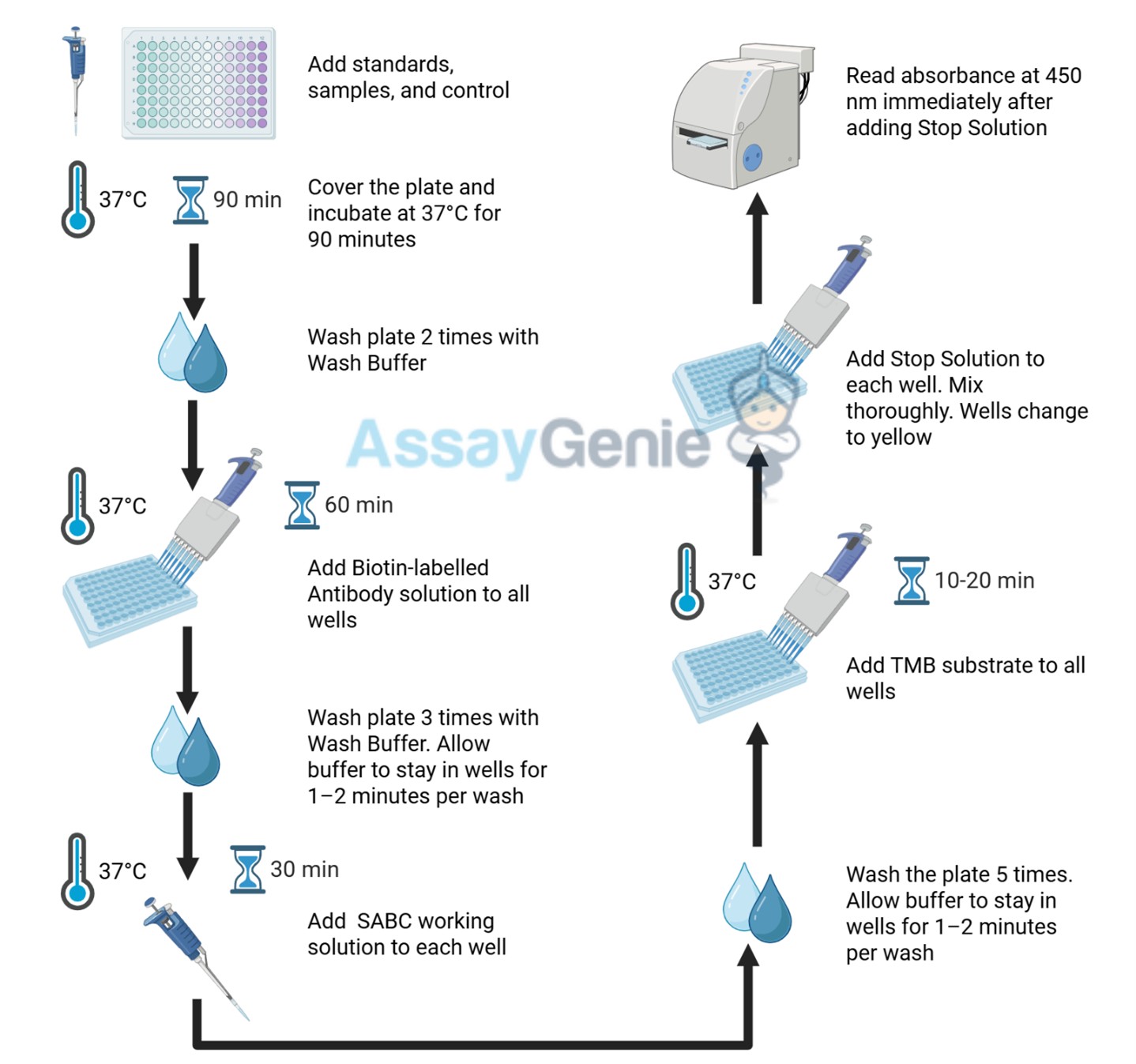
Reagent & Plate Preparation: Equilibrate reagents and TMB substrate to room temperature. Set standard, test sample and control (zero) wells on the pre-coated plate and record their positions.
2
Primary Incubation: Prepare standards, samples, blanks and load into designated wells. Incubate plate at 37°C for 90 minutes to allow antigen binding.
3
Detection Antibody Binding: Add biotin-labeled detection antibody and incubate at 37°C for 60 minutes.
4
HRP-Streptavidin Binding: Add HRP-Streptavidin (SABC) and incubate at 37°C for 30 minutes.
5
Color Development: Add TMB substrate and incubate in the dark for 10–20 minutes.
6
Stop Reaction & Reading: Add stop solution and measure absorbance at 450 nm immediately.
Sample Type
Protocol
Serum
Allow blood to clot, centrifuge at 1000 × g for 20 minutes, collect supernatant supernatant and store appropriately.
Plasma
Collect using anticoagulant tubes, centrifuge at 1000 × g for 15 minutes at 2–8°C and collect plasma.
Tissue Homogenate
Homogenize tissue in PBS with protease inhibitors, centrifuge and collect supernatant.
Cell Culture Supernatant
Centrifuge at 2500 rpm for 5 minutes and collect clarified supernatant.
Cell Lysate
Lyse cells using lysis buffer with protease inhibitors, centrifuge and collect protein supernatant.
Other Sample Types
For more information about how to process other sample types, (e.g., body fluids, breast milk & more), please contact our Tech Support Team at techsupport@assaygenie.com.
Component
Quantity
Storage
48T
96T
ELISA Microplate (Dismountable)
8×6
8×12
Place the test strips into a sealed foil bag with the desiccant. Store for 1 month at 2-8°C; Store for 12 months at -20°C.
Lyophilized Standard
1 vial
2 vial
Place the standards into a sealed foil bag with the desiccant. Store for 1 month at 2-8°C; Store for 12 months at -20°C.

 ELISA Kit (HUFI03481)")
 ELISA Kit (HUFI03481)")

 ELISA Kit (AEKE11549)")
 ELISA (Small Sample Volume) (AEKE11550)")
")
")

")