The HUS1 Antibody (CAB5407) is a high-quality antibody developed for reliable detection and analysis of target proteins. This antibody, produced in rabbits, exhibits high specificity and sensitivity towards human samples and is validated for use in Western blotting applications. By specifically binding to the HUS1 protein, this antibody allows for accurate detection and analysis in various cell types, making it a valuable asset for research in genetics, molecular biology, and cancer studies.HUS1 is a crucial factor in maintaining genomic stability and integrity by participating in cell cycle checkpoints and DNA repair mechanisms.
This antibody is validated for use in WB, IF/ICC, IP, ELISA applications and has demonstrated reactivity against Human, Mouse samples.
Product Name:
HUS1 Antibody
SKU:
CAB5407
Size:
20μL, 100μL
Reactivity:
Human, Mouse
Conjugate:
Unconjugated
Immunogen:
Recombinant protein (or fragment).This information is considered to be commercially sensitive.
0.5μg-4μg antibody for 200μg-400μg extracts of whole cells
ELISA
Recommended starting concentration is 1 μg/mL. Please optimize the concentration based on your specific assay requirements.
Synonyms:
hHUS1, HUS1
Positive Sample:
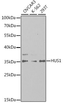
OVCAR-3, K-562, 293T
Cellular Localization:
Cytoplasm, Nucleus.
Calculated MW:
32kDa
Observed MW:
35kDa
The protein encoded by this gene is a component of an evolutionarily conserved, genotoxin-activated checkpoint complex that is involved in the cell cycle arrest in response to DNA damage. This protein forms a heterotrimeric complex with checkpoint proteins RAD9 and RAD1. In response to DNA damage, the trimeric complex interacts with another protein complex consisting of checkpoint protein RAD17 and four small subunits of the replication factor C (RFC), which loads the combined complex onto the chromatin. The DNA damage induced chromatin binding has been shown to depend on the activation of the checkpoint kinase ATM, and is thought to be an early checkpoint signaling event. Alternative splicing results in multiple transcript variants.
Purification Method
Affinity purification
Gene ID
3364
RRID
AB_2766215
Buffer Information
Store at -20℃. Avoid freeze / thaw cycles. Buffer: PBS containing 50% glycerol, preserved with proclin300 or sodium azide, pH 7.3.
Western blot analysis of various lysates using HUS1 Rabbit pAb (CAB5407) at 1:1000 dilution. Secondary antibody: HRP-conjugated Goat anti-Rabbit IgG (H+L) (CABS014) at 1:10000 dilution. Lysates/proteins: 25μg per lane. Blocking buffer: 3% nonfat dry milk in TBST. Detection: ECL Basic Kit (AbGn00020). Exposure time: 180s.
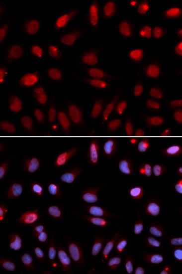
Immunofluorescence analysis of U2OS cells using HUS1 Rabbit pAb (CAB5407). Secondary antibody: Cy3-conjugated Goat anti-Rabbit IgG (H+L) (CABS007) at 1:500 dilution. Blue: DAPI for nuclear staining.
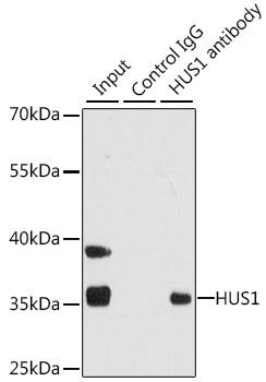
Immunoprecipitation analysis of 200 μg extracts of 293T cells using 1 μg HUS1 antibody (CAB5407). Western blot was performed from the immunoprecipitate using HUS1 antibody (CAB5407) at a dilution of 1:1000.