The ICAM3 Antibody (CAB3923) is a high-quality antibody developed for reliable detection and analysis of target proteins. This antibody, produced in rabbits, displays high specificity for human samples and has been validated for use in Western blot applications. By binding to the ICAM3 protein, it enables precise detection and analysis in various cell types, making it ideal for investigations in immunology and inflammatory diseases.ICAM3, also known as intercellular adhesion molecule 3, plays a crucial role in immune response regulation by facilitating interactions between immune cells and promoting immune cell activation.
This antibody is validated for use in WB, ELISA applications and has demonstrated reactivity against Human samples.
Product Name:
ICAM3 Antibody
SKU:
CAB3923
Size:
20μL, 100μL
Reactivity:
Human
Conjugate:
Unconjugated
Immunogen:
Recombinant protein (or fragment).This information is considered to be commercially sensitive.
Recommended starting concentration is 1 μg/mL. Please optimize the concentration based on your specific assay requirements.
Synonyms:
CD50, CDW50, ICAM-R, ICAM3
Positive Sample:
U-937
Cellular Localization:
Membrane, Single-Pass Type I Membrane Protein.
Calculated MW:
60kDa
Observed MW:
140kDa
The protein encoded by this gene is a member of the intercellular adhesion molecule (ICAM) family. All ICAM proteins are type I transmembrane glycoproteins, contain 2-9 immunoglobulin-like C2-type domains, and bind to the leukocyte adhesion LFA-1 protein. This protein is constitutively and abundantly expressed by all leucocytes and may be the most important ligand for LFA-1 in the initiation of the immune response. It functions not only as an adhesion molecule, but also as a potent signalling molecule. Alternative splicing results in multiple transcript variants encoding different isoforms.
Purification Method
Affinity purification
Gene ID
3385
RRID
AB_2765389
Buffer Information
Store at -20℃. Avoid freeze / thaw cycles. Buffer: PBS containing 50% glycerol, preserved with proclin300 or sodium azide, pH 7.3.
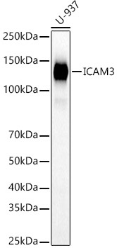
Western blot analysis of lysates from U-937 cells, using ICAM3 Rabbit pAb (CAB3923) at 1:1000 dilution. Secondary antibody: HRP-conjugated Goat anti-Rabbit IgG (H+L) (CABS014) at 1:10000 dilution. Lysates/proteins: 25μg per lane. Blocking buffer: 3% nonfat dry milk in TBST. Detection: ECL Basic Kit (AbGn00020). Exposure time: 10s.