The IDO1 Antibody (CAB1614) is a high-quality antibody developed for reliable detection and analysis of target proteins. This antibody, generated in rabbits, is highly specific for human samples and has been validated for use in Western blot applications. By binding to the IDO1 protein, this antibody allows for the detection and analysis of IDO1 expression in various cell types, making it ideal for studies in immunology, cancer research, and autoimmune disorders.IDO1, also known as indoleamine 2,3-dioxygenase 1, plays a crucial role in immune escape mechanisms utilized by cancer cells and other immune-mediated diseases.
This antibody is validated for use in WB, IF/ICC, ELISA applications and has demonstrated reactivity against Human, Rat samples.
Product Name:
IDO1 Antibody
SKU:
CAB1614
Size:
20μL, 100μL
Reactivity:
Human, Rat
Conjugate:
Unconjugated
Immunogen:
Recombinant protein (or fragment).This information is considered to be commercially sensitive.
Recommended starting concentration is 1 μg/mL. Please optimize the concentration based on your specific assay requirements.
Synonyms:
IDO, INDO, IDO-1, IDO1
Positive Sample:
SKOV3, A-549 treated with hIFN-γ
Cellular Localization:
Cytoplasm, Cytosol.
Calculated MW:
45kDa
Observed MW:
43kDa
This gene encodes indoleamine 2,3-dioxygenase (IDO) - a heme enzyme that catalyzes the first and rate-limiting step in tryptophan catabolism to N-formyl-kynurenine. This enzyme acts on multiple tryptophan substrates including D-tryptophan, L-tryptophan, 5-hydroxy-tryptophan, tryptamine, and serotonin. This enzyme is thought to play a role in a variety of pathophysiological processes such as antimicrobial and antitumor defense, neuropathology, immunoregulation, and antioxidant activity. Through its expression in dendritic cells, monocytes, and macrophages this enzyme modulates T-cell behavior by its peri-cellular catabolization of the essential amino acid tryptophan.
Purification Method
Affinity purification
Gene ID
3620
RRID
AB_2763586
Buffer Information
Store at -20℃. Avoid freeze / thaw cycles. Buffer: PBS with 0.09% Sodium azide,50% glycerol,pH7.3.
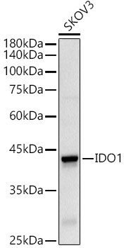
Western blot analysis of lysates from SKOV3 cells, using IDO1 Rabbit pAb (CAB1614) at 1:500 dilution. Secondary antibody: HRP-conjugated Goat anti-Rabbit IgG (H+L) (CABS014) at 1:10000 dilution. Lysates/proteins: 25μg per lane. Blocking buffer: 3% nonfat dry milk in TBST. Detection: ECL Basic Kit (AbGn00020). Exposure time: 90s.
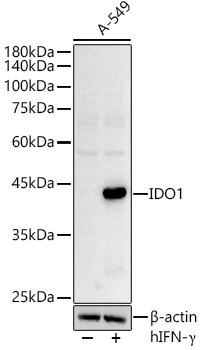
Western blot analysis of lysates from A-549 cells, using IDO1 Rabbit pAb (CAB1614) at 1:500 dilution. A-549 cells were treated with hIFN-γ(100 ng/mL) at 37℃ for 48 hours. Secondary antibody: HRP-conjugated Goat anti-Rabbit IgG (H+L) (CABS014) at 1:10000 dilution. Lysates/proteins: 25μg per lane. Blocking buffer: 3% nonfat dry milk in TBST. Detection: ECL Basic Kit (AbGn00020). Exposure time: 90s.