The IFIH1 Antibody (CAB13645) is a high-quality antibody developed for reliable detection and analysis of target proteins. This antibody, produced in rabbits, is highly specific to human IFIH1 and has been validated for use in Western blot applications. By targeting the IFIH1 protein, this antibody enables researchers to detect and analyze IFIH1 expression in various cell types, providing valuable insights into the role of IFIH1 in immune signaling pathways.IFIH1, also known as MDA5 (melanoma differentiation-associated protein 5), is a key player in antiviral immunity, sensing viral RNA and triggering immune responses to combat viral infections.
This antibody is validated for use in WB, IHC-P, IF/ICC, ELISA applications and has demonstrated reactivity against Human, Mouse, Rat samples.
Product Name:
IFIH1 Antibody
SKU:
CAB13645
Size:
20μL, 100μL
Reactivity:
Human, Mouse, Rat
Conjugate:
Unconjugated
Immunogen:
Recombinant protein (or fragment).This information is considered to be commercially sensitive.
IFIH1 encodes MDA5 which is an intracellular sensor of viral RNA that triggers the innate immune response. Sensing RNA length and secondary structure, MDA5 binds dsRNA oligonucleotides with a modified DExD/H-box helicase core and a C-terminal domain, thus leading to a proinflammatory response that includes interferons. It has been shown that Coronaviruses (CoVs) as well as various other virus families, are capable of evading the MDA5-dependent interferon response, thus impeding the activation of the innate immune response to infection. MDA5 has also been shown to play an important role in enhancing natural killer cell function in malaria infection. In addition to its protective role in antiviral responses, MDA5 has been implicated in autoimmune and autoinflammatory diseases such as type 1 diabetes, systemic lupus erythematosus, and Aicardi-Goutieres syndrome
Purification Method
Affinity purification
Gene ID
64135
RRID
AB_2760507
Buffer Information
Store at -20℃. Avoid freeze / thaw cycles. Buffer: PBS with 0.09% Sodium azide,50% glycerol,pH7.3.
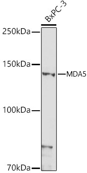
Western blot analysis of lysates from BxPC-3 cells, using MDA5 Rabbit pAb (CAB13645) at 1:1000 dilution. Secondary antibody: HRP-conjugated Goat anti-Rabbit IgG (H+L) (CABS014) at 1:10000 dilution. Lysates/proteins: 25 μg per lane. Blocking buffer: 3% nonfat dry milk in TBST. Detection: ECL Basic Kit (AbGn00020). Exposure time: 180 s.
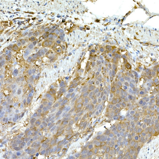
Immunohistochemistry analysis of paraffin-embedded Human esophageal cancer using MDA5 Rabbit pAb (CAB13645) at dilution of 1:50 (40x lens). High pressure antigen retrieval performed with 0.01M Citrate buffer (pH 6.0) prior to IHC staining.
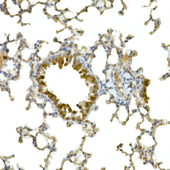
Immunohistochemistry analysis of paraffin-embedded Mouse lung using MDA5 Rabbit pAb (CAB13645) at dilution of 1:50 (40x lens). High pressure antigen retrieval performed with 0.01M Citrate buffer (pH 6.0) prior to IHC staining.
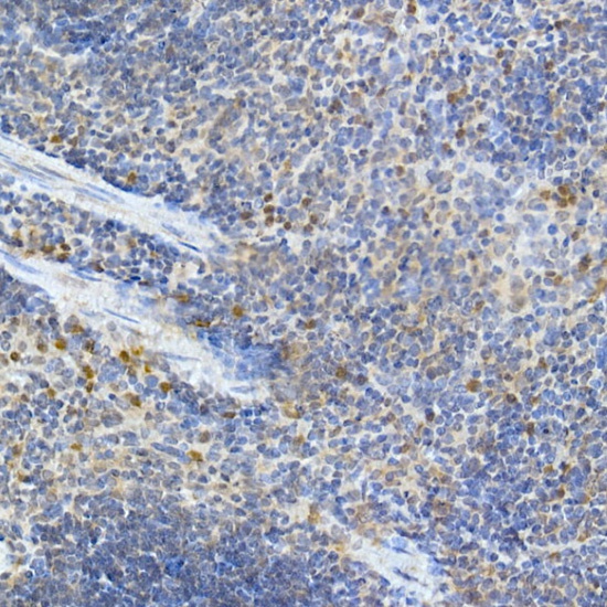
Immunohistochemistry analysis of paraffin-embedded Mouse spleen using MDA5 Rabbit pAb (CAB13645) at dilution of 1:50 (40x lens). High pressure antigen retrieval performed with 0.01M Citrate buffer (pH 6.0) prior to IHC staining.
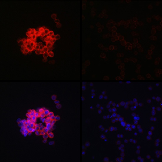
Immunofluorescence analysis of THP-1 treated with LPS(32h) and THP-1 cells using MDA5 Rabbit pAb (CAB13645) at dilution of 1:100 (40x lens). Secondary antibody: Cy3-conjugated Goat anti-Rabbit IgG (H+L) (CABS007) at 1:500 dilution. Blue: DAPI for nuclear staining.


")


