The IGFBP1 Monoclonal Antibody (CAB11672) is a high-quality antibody developed for reliable detection and analysis of target proteins. This antibody, derived from rabbits, has been validated for use in various research applications, including Western blotting and immunohistochemistry.IGFBP1, also known as insulin-like growth factor-binding protein 1, is a key player in the growth hormone axis and has been implicated in various physiological processes, such as cell proliferation, differentiation, and survival. Dysregulation of IGFBP1 has been associated with conditions like diabetes, cardiovascular disease, and cancer, making it a promising target for therapeutic intervention.
This antibody is validated for use in WB, ELISA applications and has demonstrated reactivity against Human samples.
Product Name:
IGFBP1 Monoclonal Antibody
SKU:
CAB11672
Size:
20μL, 100μL
Reactivity:
Human
Clone Number:
ARC0671
Conjugate:
Unconjugated
Immunogen:
Synthetic peptide. This information is considered to be commercially sensitive.
Recommended starting concentration is 1 μg/mL. Please optimize the concentration based on your specific assay requirements.
Synonyms:
AFBP, IBP1, PP12, IGF-BP25, hIGFBP-1, IGFBP1
Positive Sample:
Hep G2
Cellular Localization:
Secreted.
Calculated MW:
28kDa
Observed MW:
30kDa
This gene is a member of the insulin-like growth factor binding protein (IGFBP) family and encodes a protein with an IGFBP N-terminal domain and a thyroglobulin type-I domain. The encoded protein, mainly expressed in the liver, circulates in the plasma and binds both insulin-like growth factors (IGFs) I and II, prolonging their half-lives and altering their interaction with cell surface receptors. This protein is important in cell migration and metabolism. Low levels of this protein may be associated with impaired glucose tolerance, vascular disease and hypertension in human patients.
Purification Method
Affinity purification
Gene ID
3484
RRID
AB_2861625
Buffer Information
Store at -20℃. Avoid freeze / thaw cycles. Buffer: PBS containing 50% glycerol and 0.05% BSA, preserved with proclin300 or sodium azide, pH 7.3.
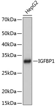
Western blot analysis of lysates from HepG2 cells, using IGFBP1 Rabbit mAb (CAB11672) at 1:1000 dilution. Secondary antibody: HRP-conjugated Goat anti-Rabbit IgG (H+L) (CABS014) at 1:10000 dilution. Lysates/proteins: 25μg per lane. Blocking buffer: 3% nonfat dry milk in TBST. Detection: ECL Basic Kit (AbGn00020). Exposure time: 1s.