The IGFBP5 Antibody (CAB12451) is a high-quality antibody developed for reliable detection and analysis of target proteins. This antibody, produced in rabbits, exhibits high reactivity with human samples and has been validated for use in Western blot applications. By specifically binding to IGFBP5, it enables precise detection and analysis of this protein in various cell types, making it well-suited for investigations in the fields of molecular biology and cancer research.
This antibody is validated for use in WB, IHC-P, IF/ICC, ELISA applications and has demonstrated reactivity against Human, Mouse, Rat samples.
Product Name:
IGFBP5 Antibody
SKU:
CAB12451
Size:
20μL, 100μL
Reactivity:
Human, Mouse, Rat
Conjugate:
Unconjugated
Immunogen:
Recombinant protein (or fragment).This information is considered to be commercially sensitive.
Recommended starting concentration is 1 μg/mL. Please optimize the concentration based on your specific assay requirements.
Synonyms:
IBP5, IGFBP5
Positive Sample:
Mouse kidney
Cellular Localization:
Secreted.
Calculated MW:
31kDa
Observed MW:
33kDa
Enables insulin-like growth factor I binding activity. Involved in several processes, including cellular response to cAMP; regulation of smooth muscle cell migration; and regulation of smooth muscle cell proliferation. Part of insulin-like growth factor ternary complex. Biomarker of pulmonary fibrosis.
Purification Method
Affinity purification
Gene ID
3488
RRID
AB_2759295
Buffer Information
Store at -20℃. Avoid freeze / thaw cycles. Buffer: PBS containing 50% glycerol, preserved with proclin300 or sodium azide, pH 7.3.
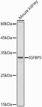
Western blot analysis of lysates from Mouse kidney, using IGFBP5 Rabbit pAb (CAB12451) at 1:500 dilution. Secondary antibody: HRP-conjugated Goat anti-Rabbit IgG (H+L) (CABS014) at 1:10000 dilution. Lysates/proteins: 25μg per lane. Blocking buffer: 3% nonfat dry milk in TBST. Detection: ECL Basic Kit (AbGn00020). Exposure time: 30s.
Western blot analysis of lysates from Mouse uterus, using IGFBP5 Rabbit pAb (CAB12451) at 1:1000 dilution. Secondary antibody: HRP-conjugated Goat anti-Rabbit IgG (H+L) (CABS014) at 1:10000 dilution. Lysates/proteins: 25μg per lane. Blocking buffer: 3% nonfat dry milk in TBST. Detection: ECL Basic Kit (AbGn00020). Exposure time: 90s.
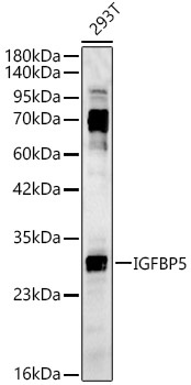
Western blot analysis of lysates from 293T cells, using IGFBP5 Rabbit pAb (CAB12451) at 1:2000 dilution. Secondary antibody: HRP-conjugated Goat anti-Rabbit IgG (H+L) (CABS014) at 1:10000 dilution. Lysates/proteins: 25μg per lane. Blocking buffer: 3% nonfat dry milk in TBST. Detection: ECL Basic Kit (AbGn00020). Exposure time: 45s.
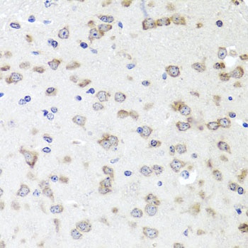
Immunohistochemistry analysis of paraffin-embedded Mouse brain using IGFBP5 Rabbit pAb (CAB12451) at dilution of 1:100 (40x lens). Microwave antigen retrieval performed with 0.01M PBS Buffer (pH 7.2) prior to IHC staining.
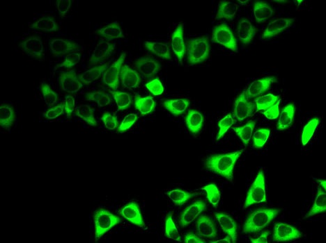
Immunofluorescence analysis of HeLa cells using IGFBP5 Rabbit pAb (CAB12451).Secondary antibody: Cy3-conjugated Goat anti-Rabbit IgG (H+L) (CABS007) at 1:500 dilution.
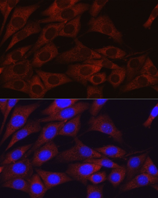
Immunofluorescence analysis of NIH/3T3 cells using IGFBP5 Rabbit pAb (CAB12451) at dilution of 1:100 (40x lens). Secondary antibody: Cy3-conjugated Goat anti-Rabbit IgG (H+L) (CABS007) at 1:500 dilution. Blue: DAPI for nuclear staining.
Immunofluorescence analysis of PC-12 cells using IGFBP5 Rabbit pAb (CAB12451) at dilution of 1:100 (40x lens). Secondary antibody: Cy3-conjugated Goat anti-Rabbit IgG (H+L) (CABS007) at 1:500 dilution. Blue: DAPI for nuclear staining.