The IGFBP7 Monoclonal Antibody (CAB4615) is a high-quality antibody developed for reliable detection and analysis of target proteins. This antibody, developed using rabbit monoclonal technology, exhibits high specificity and sensitivity for detecting IGFBP7 in human samples.IGFBP7 is involved in regulating cell proliferation and survival, making it a potential target for cancer research and therapeutic development. By targeting IGFBP7, researchers can gain insights into its function in promoting or inhibiting tumor growth, providing valuable information for the development of targeted cancer therapies.
This antibody is validated for use in WB, IF/ICC, ELISA applications and has demonstrated reactivity against Human, Mouse, Rat samples.
Product Name:
IGFBP7 Monoclonal Antibody
SKU:
CAB4615
Size:
20μL, 100μL
Reactivity:
Human, Mouse, Rat
Clone Number:
ARC1052
Conjugate:
Unconjugated
Immunogen:
Synthetic peptide. This information is considered to be commercially sensitive.
This gene encodes a member of the insulin-like growth factor (IGF)-binding protein (IGFBP) family. IGFBPs bind IGFs with high affinity, and regulate IGF availability in body fluids and tissues and modulate IGF binding to its receptors. This protein binds IGF-I and IGF-II with relatively low affinity, and belongs to a subfamily of low-affinity IGFBPs. It also stimulates prostacyclin production and cell adhesion. Alternatively spliced transcript variants encoding different isoforms have been described for this gene, and one variant has been associated with retinal arterial macroaneurysm (PMID:21835307).
Purification Method
Affinity purification
Gene ID
3490
RRID
AB_2863310
Buffer Information
Store at -20℃. Avoid freeze / thaw cycles. Buffer: PBS containing 50% glycerol and 0.05% BSA, preserved with proclin300 or sodium azide, pH 7.3.
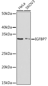
Western blot analysis of various lysates using IGFBP7 Rabbit mAb (CAB4615) at 1:1000 dilution. Secondary antibody: HRP-conjugated Goat anti-Rabbit IgG (H+L) (CABS014) at 1:10000 dilution. Lysates/proteins: 25μg per lane. Blocking buffer: 3% nonfat dry milk in TBST. Detection: ECL Basic Kit (AbGn00020). Exposure time: 90s.
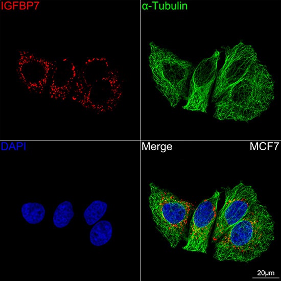
Confocal imaging of MCF7 cells using IGFBP7 Rabbit mAb (CAB4615,at dilution of 1:100) (Red). The cells were counterstained with α-Tubulin Mouse mAb (AC012,dilution 1:400) (Green). DAPI was used for nuclear staining (blue). Objective: 100x.


")


