The IKBKAP Antibody (CAB10127) is a high-quality antibody developed for reliable detection and analysis of target proteins. This antibody, produced in rabbits, exhibits high reactivity with human samples and has been validated for use in Western blot applications. By binding to the IKBKAP protein, this antibody enables the detection and analysis of IKBKAP in various cell types, making it ideal for investigations in immunology and inflammation-related research.IKBKAP, also known as inhibitor of kappa light polypeptide gene enhancer in B-cells, kinase complex-associated protein, plays a crucial role in the NF-kappa-B signaling pathway, which is involved in the regulation of immune and inflammatory responses.
This antibody is validated for use in WB, IHC-P, ELISA applications and has demonstrated reactivity against Human, Mouse, Rat samples.
Product Name:
IKBKAP Antibody
SKU:
CAB10127
Size:
20μL, 100μL
Reactivity:
Human, Mouse, Rat
Conjugate:
Unconjugated
Immunogen:
Recombinant protein (or fragment).This information is considered to be commercially sensitive.
Recommended starting concentration is 1 μg/mL. Please optimize the concentration based on your specific assay requirements.
Synonyms:
FD, DYS, IKAP, IKI3, TOT1, IKBKAP
Positive Sample:
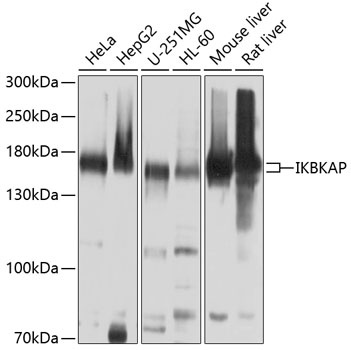
HeLa, HepG2, U-251MG, HL-60, Mouse liver, Rat liver
Cellular Localization:
Cytoplasm, Nucleus.
Calculated MW:
150kDa
Observed MW:
150kDa
The protein encoded by this gene is a scaffold protein and a regulator for three different kinases involved in proinflammatory signaling. The encoded protein can bind NF-kappa-B-inducing kinase and I-kappa-B kinases through separate domains and assemble them into an active kinase complex. Mutations in this gene have been associated with familial dysautonomia. Alternative splicing results in multiple transcript variants encoding different isoforms.
Purification Method
Affinity purification
Gene ID
8518
RRID
AB_2757652
Buffer Information
Store at -20℃. Avoid freeze / thaw cycles. Buffer: PBS containing 50% glycerol, preserved with proclin300 or sodium azide, pH 7.3.
Western blot analysis of various lysates using IKBKAP Rabbit pAb (CAB10127) at 1:1000 dilution. Secondary antibody: HRP-conjugated Goat anti-Rabbit IgG (H+L) (CABS014) at 1:10000 dilution. Lysates/proteins: 25μg per lane. Blocking buffer: 3% nonfat dry milk in TBST. Detection: ECL Basic Kit (AbGn00020). Exposure time: 10s.
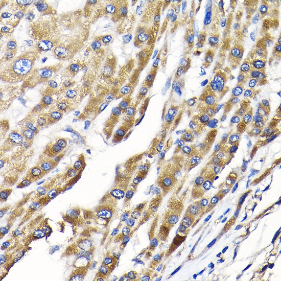
Immunohistochemistry analysis of paraffin-embedded Human liver cancer using IKBKAP Rabbit pAb (CAB10127) at dilution of 1:100 (40x lens). High pressure antigen retrieval performed with 0.01M Citrate buffer (pH 6.0) prior to IHC staining.
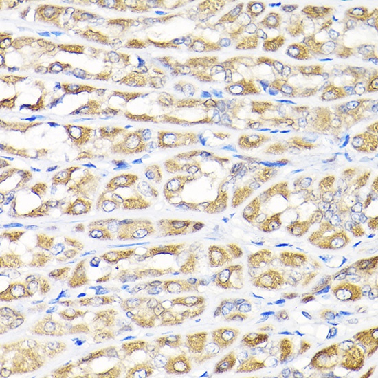
Immunohistochemistry analysis of paraffin-embedded Rat stomach using IKBKAP Rabbit pAb (CAB10127) at dilution of 1:100 (40x lens). High pressure antigen retrieval performed with 0.01M Citrate buffer (pH 6.0) prior to IHC staining.