The IL-1 alpha Monoclonal Antibody (CAB22766) is a high-quality antibody developed for reliable detection and analysis of target proteins. This monoclonal antibody, developed using cutting-edge technology, is ideal for use in a range of applications including ELISA, Western blot, and immunohistochemistry.IL-1 Alpha is a key player in the regulation of immune responses and inflammation, making it a crucial target for studying diseases such as arthritis, autoimmune disorders, and cancer.
This antibody is validated for use in WB, ELISA applications and has demonstrated reactivity against Mouse samples.
Product Name:
IL-1 alpha Monoclonal Antibody
SKU:
CAB22766
Size:
20μL, 100μL
Reactivity:
Mouse
Clone Number:
ARC58205
Conjugate:
Unconjugated
Immunogen:
Recombinant protein (or fragment).This information is considered to be commercially sensitive.
Recommended starting concentration is 1 μg/mL. Please optimize the concentration based on your specific assay requirements.
Synonyms:
Il-1a, IL-1 alpha
Positive Sample:
Recombinant Mouse IL-1 alpha Protein, RAW 264.7 treated with LPS
Cellular Localization:
Nucleus, Secreted, Cytoplasm .
Calculated MW:
31kDa
Observed MW:
20-25kDa/31kDa
Enables cytokine activity and interleukin-1 receptor binding activity. Involved in positive regulation of angiogenesis and positive regulation of vascular endothelial growth factor production. Acts upstream of or within several processes, including cell surface receptor signaling pathway; connective tissue replacement involved in inflammatory response wound healing; and positive regulation of cytokine production. Located in cytoplasm and extracellular space. Is expressed in several structures, including alimentary system; brain; hemolymphoid system gland; reproductive system; and respiratory system. Human ortholog(s) of this gene implicated in several diseases, including Fabry disease; Schnitzler syndrome; autoimmune disease (multiple); hematologic cancer (multiple); and lung disease (multiple). Orthologous to human IL1A (interleukin 1 alpha).
Purification Method
Affinity purification
Gene ID
16175
Buffer Information
Store at -20℃. Avoid freeze / thaw cycles. Buffer: PBS containing 50% glycerol and 0.05% BSA, preserved with proclin300 or sodium azide, pH 7.3.
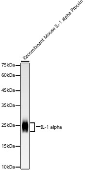
Western blot analysis of Recombinant Mouse IL-1 alpha Protein using IL-1 alpha Rabbit mAb (CAB22766) at 1:10000 dilution. Secondary antibody: HRP-conjugated Goat anti-Rabbit IgG (H+L) (CABS014) at 1:10000 dilution. Lysates/proteins: 10 ng per lane. Blocking buffer: 3% nonfat dry milk in TBST. Detection: ECL Basic Kit (AbGn00020). Exposure time: 180 s.
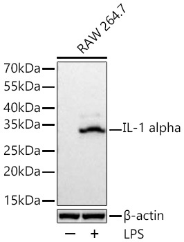
Western blot analysis of lysates from RAW 264.7 cells using IL-1 alpha Rabbit mAb (CAB22766) at 1:1000 dilution incubated at room temperature for 1.5 hours. RAW 264.7 cells were treated with LPS (1 μg/mL) for 8 hours. Secondary antibody: HRP-conjugated Goat anti-Rabbit IgG (H+L) (CABS014) at 1:10000 dilution. Lysates/proteins: 30 μg per lane. Blocking buffer: 3% nonfat dry milk in TBST. Detection: ECL Basic Kit (AbGn00020). Exposure time: 5 s.
 at 1:10000 dilution. Secondary antibody: HRP Goat Anti-Rabbit IgG (H+L) at 1:10000 dilution. Lysates/proteins: 25μg per lane. Blocking buffer: 3% nonfat dry milk in TBST.")
 at 1:10000 dilution. Secondary antibody: HRP Goat Anti-Rabbit IgG (H+L) at 1:10000 dilution. Lysates/proteins: 25μg per lane. Blocking buffer: 3% nonfat dry milk in TBST.")

