The IL1beta Monoclonal Antibody (CAB22257) is a high-quality antibody developed for reliable detection and analysis of target proteins. This antibody, developed using cutting-edge technology, is highly specific for IL-1 and is ideal for use in various immunoassays and experimental techniques.IL-1 is a key player in the initiation and regulation of immune responses, making it a crucial focus in studies related to inflammatory diseases, autoimmune disorders, and cancer.
This antibody is validated for use in WB, IF/ICC, ELISA applications and has demonstrated reactivity against Mouse samples.
Product Name:
IL1beta Monoclonal Antibody
SKU:
CAB22257
Size:
20μL, 100μL
Reactivity:
Mouse
Clone Number:
ARC56180
Conjugate:
Unconjugated
Immunogen:
Recombinant protein (or fragment).This information is considered to be commercially sensitive.
The protein encoded by this gene is a member of the interleukin 1 cytokine family. This cytokine is produced by activated macrophages as a proprotein, which is proteolytically processed to its active form by caspase 1. The encoded protein plays a role in thymocyte proliferation and is involved in the inflammatory response.
Purification Method
Affinity purification
Gene ID
16176
Buffer Information
Store at -20℃. Avoid freeze / thaw cycles. Buffer: PBS containing 50% glycerol and 0.05% BSA, preserved with proclin300 or sodium azide, pH 7.3.
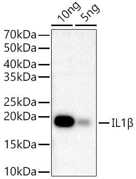
Western blot analysis of Recombinant Mouse IL-1 beta Protein (RP01340), using IL1β Rabbit mAb (CAB22257) at1:2000 dilution. Secondary antibody: HRP-conjugated Goat anti-Rabbit IgG (H+L) (CABS014) at 1:10000 dilution. Lysates/proteins: 10ng/5ng per lane. Blocking buffer: 3% nonfat dry milk in TBST. Detection: ECL Basic Kit (AbGn00020). Exposure time: 20 s.
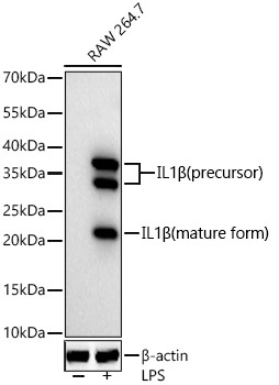
Western blot analysis of lysates from RAW 264.7 cells using IL1β Rabbit mAb (CAB22257) at 1:1000 dilution incubated overnight at 4℃. Raw264.7 cells were treated with LPS (1 μg/ml) at 37℃ for 8 hours. Secondary antibody: HRP-conjugated Goat anti-Rabbit IgG (H+L) (CABS014) at 1:10000 dilution. Lysates/proteins: 30 μg per lane. Blocking buffer: 3% nonfat dry milk in TBST. Detection: ECL Basic Kit (AbGn00020). Exposure time: 90 s.
 at1:2000 dilution. Secondary antibody: HRP Goat Anti-Rabbit IgG (H+L) at 1:10000 dilution. Lysates/proteins: 25μg per lane. Blocking buffer: 3% nonfat dry milk in TBST.")
 at1:2000 dilution. Secondary antibody: HRP Goat Anti-Rabbit IgG (H+L) at 1:10000 dilution. Lysates/proteins: 25μg per lane. Blocking buffer: 3% nonfat dry milk in TBST.")


