The IL17C Antibody (CAB10587) is a high-quality antibody developed for reliable detection and analysis of target proteins. This antibody, produced in rabbits, is highly specific for IL-17C in human samples and is suitable for use in Western blot experiments. By binding to IL-17C, it enables researchers to detect and analyze the protein in various cell types, making it an essential reagent for studies in immunology and inflammatory diseases.IL-17C is known for its involvement in promoting inflammation and immune responses, making it a potential target for therapeutic interventions in conditions such as autoimmune diseases and inflammatory disorders.
This antibody is validated for use in WB, IF/ICC, ELISA applications and has demonstrated reactivity against Human, Mouse, Rat samples.
Product Name:
IL17C Antibody
SKU:
CAB10587
Size:
20μL, 100μL
Reactivity:
Human, Mouse, Rat
Conjugate:
Unconjugated
Immunogen:
Recombinant protein (or fragment).This information is considered to be commercially sensitive.
Recommended starting concentration is 1 μg/mL. Please optimize the concentration based on your specific assay requirements.
Synonyms:
CX2, IL-17C, IL17C
Positive Sample:
A549
Cellular Localization:
Secreted.
Calculated MW:
22kDa
Observed MW:
22kDa/18kDa
The protein encoded by this gene is a T cell-derived cytokine that shares the sequence similarity with IL17. This cytokine was reported to stimulate the release of tumor necrosis factor alpha and interleukin 1 beta from a monocytic cell line. The expression of this cytokine was found to be restricted to activated T cells.
Purification Method
Affinity purification
Gene ID
27189
RRID
AB_2758128
Buffer Information
Store at -20℃. Avoid freeze / thaw cycles. Buffer: PBS containing 50% glycerol, preserved with proclin300 or sodium azide, pH 7.3.
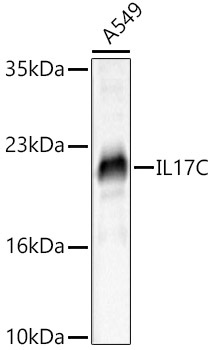
Western blot analysis of lysates from A549 cells , using IL17C Rabbit pAb (CAB10587) at 1:1000 dilution. Secondary antibody: HRP-conjugated Goat anti-Rabbit IgG (H+L) (CABS014) at 1:10000 dilution. Lysates/proteins: 25μg per lane. Blocking buffer: 3% nonfat dry milk in TBST. Detection: ECL Basic Kit (AbGn00020). Exposure time: 30s.
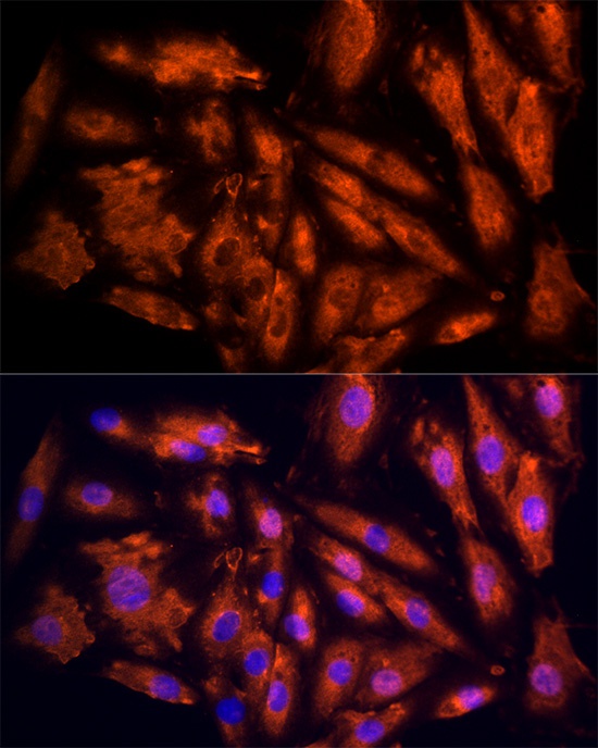
Immunofluorescence analysis of H9C2 cells using IL17C Rabbit pAb (CAB10587) at dilution of 1:100 (40x lens). Secondary antibody: Cy3-conjugated Goat anti-Rabbit IgG (H+L) (CABS007) at 1:500 dilution. Blue: DAPI for nuclear staining.
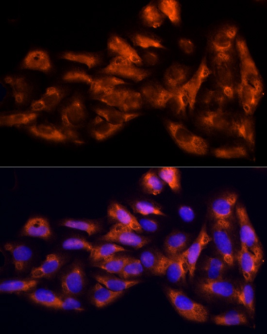
Immunofluorescence analysis of U2OS cells using IL17C Rabbit pAb (CAB10587) at dilution of 1:100 (40x lens). Secondary antibody: Cy3-conjugated Goat anti-Rabbit IgG (H+L) (CABS007) at 1:500 dilution. Blue: DAPI for nuclear staining.