The IMMT Antibody (CAB2751) is a high-quality antibody developed for reliable detection and analysis of target proteins. This rabbit-raised antibody is highly specific to human samples and has been validated for use in Western blot applications. By targeting the IMM-T protein, this antibody enables researchers to detect and analyze IMM-T levels in various cell types, making it ideal for studies in metabolism, bioenergetics, and mitochondrial disorders.IMMT, also known as inner membrane protein, plays a crucial role in maintaining mitochondrial integrity and function. Dysfunction in the IMM-T gene has been linked to various metabolic and mitochondrial diseases, making it an important target for research in the fields of cell biology and metabolism.
This antibody is validated for use in WB, IHC-P, IF/ICC, ELISA applications and has demonstrated reactivity against Human, Mouse, Rat samples.
Product Name:
IMMT Antibody
SKU:
CAB2751
Size:
20μL, 100μL
Reactivity:
Human, Mouse, Rat
Conjugate:
Unconjugated
Immunogen:
Recombinant protein (or fragment).This information is considered to be commercially sensitive.
Enables RNA binding activity. Involved in cristae formation. Located in mitochondrial inner membrane. Part of MICOS complex.
Purification Method
Affinity purification
Gene ID
10989
RRID
AB_2764600
Buffer Information
Store at -20℃. Avoid freeze / thaw cycles. Buffer: PBS containing 50% glycerol, preserved with proclin300 or sodium azide, pH 7.3.
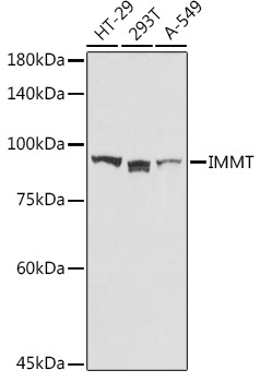
Western blot analysis of various lysates using IMMT Rabbit pAb (CAB2751) at 1:1000 dilution. Secondary antibody: HRP-conjugated Goat anti-Rabbit IgG (H+L) (CABS014) at 1:10000 dilution. Lysates/proteins: 25μg per lane. Blocking buffer: 3% nonfat dry milk in TBST. Detection: ECL Basic Kit (AbGn00020). Exposure time: 30s.
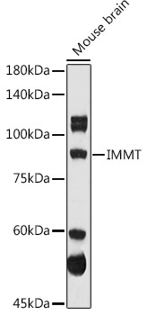
Western blot analysis of lysates from Mouse brain, using IMMT Rabbit pAb (CAB2751) at 1:1000 dilution. Secondary antibody: HRP-conjugated Goat anti-Rabbit IgG (H+L) (CABS014) at 1:10000 dilution. Lysates/proteins: 25μg per lane. Blocking buffer: 3% nonfat dry milk in TBST. Detection: ECL Enhanced Kit (AbGn00021). Exposure time: 180s.
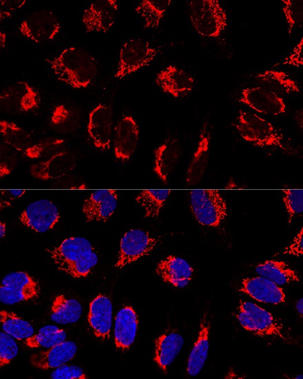
Confocal immunofluorescence analysis of U2OS cells using IMMT Rabbit pAb (CAB2751) at dilution of 1:100. Blue: DAPI for nuclear staining.