Inhibin beta A (INHBA) Monoclonal Antibody (CAB5232)
The Inhibin beta A (INHBA) Monoclonal Antibody (CAB5232) is a high-quality antibody developed for reliable detection and analysis of target proteins. This antibody, generated in rabbits, is optimized for recognition of human samples and is validated for use in various applications, including Western blotting and immunohistochemistry.Inhibin Beta A is a crucial regulator of cell proliferation and differentiation, with implications in various physiological processes such as reproduction and inflammation. Its role in modulating immune responses makes it a promising target for studies in immunology, cancer research, and reproductive biology.
This antibody is validated for use in WB, IF/ICC, ELISA applications and has demonstrated reactivity against Human, Mouse, Rat samples.
Product Name:
Inhibin beta A (INHBA) Monoclonal Antibody
SKU:
CAB5232
Size:
20μL, 100μL
Reactivity:
Human, Mouse, Rat
Clone Number:
ARC1177
Conjugate:
Unconjugated
Immunogen:
Synthetic peptide. This information is considered to be commercially sensitive.
Recommended starting concentration is 1 μg/mL. Please optimize the concentration based on your specific assay requirements.
Synonyms:
EDF, FRP, Inhibin beta A (INHBA)
Positive Sample:
SW480, Caco-2, Rat testis, Mouse ovary
Cellular Localization:
Secreted.
Calculated MW:
47kDa
Observed MW:
47kDa
This gene encodes a member of the TGF-beta (transforming growth factor-beta) superfamily of proteins. The encoded preproprotein is proteolytically processed to generate a subunit of the dimeric activin and inhibin protein complexes. These complexes activate and inhibit, respectively, follicle stimulating hormone secretion from the pituitary gland. The encoded protein also plays a role in eye, tooth and testis development. Elevated expression of this gene may be associated with cancer cachexia in human patients.
Purification Method
Affinity purification
Gene ID
3624
RRID
AB_2863495
Buffer Information
Store at -20℃. Avoid freeze / thaw cycles. Buffer: PBS containing 50% glycerol and 0.05% BSA, preserved with proclin300 or sodium azide, pH 7.3.
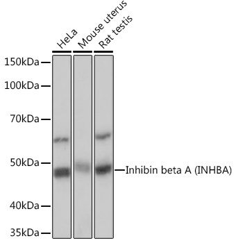
Western blot analysis of various lysates using Inhibin beta A (INHBA) (INHBA) Rabbit mAb (CAB5232) at 1:1000 dilution. Secondary antibody: HRP-conjugated Goat anti-Rabbit IgG (H+L) (CABS014) at 1:10000 dilution. Lysates/proteins: 25μg per lane. Blocking buffer: 3% nonfat dry milk in TBST. Detection: ECL Basic Kit (AbGn00020). Exposure time: 1s.
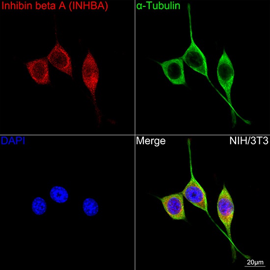
Confocal imaging of NIH/3T3 cells using Inhibin beta A (INHBA) Rabbit mAb (CAB5232, dilution 1:100) followed by a further incubation with Cy3 Goat Anti-Rabbit IgG (H+L) (CABS007, dilution 1:500) (Red). The cells were counterstained with α-Tubulin Mouse mAb (AC012, dilution 1:400) followed by incubation with ABflo® 488-conjugated Goat Anti-Mouse IgG (H+L) Ab (CABS076, dilution 1:500) (Green). DAPI was used for nuclear staining (Blue). Objective: 100x.


 CLIA Kit (MOES00384)")

