The ITLN1 Antibody (CAB7234) is a high-quality antibody developed for reliable detection and analysis of target proteins. This antibody, produced in rabbits, is highly specific to human samples and has been validated for use in various immunoassays, including Western blotting.Intelectin-1, also known as Omentin-1, is known for its role in regulating inflammation in the gut and promoting a healthy immune system. Its expression has been linked to various diseases, including inflammatory bowel diseases and colorectal cancer.
This antibody is validated for use in WB, IHC-P, ELISA, IF-P applications and has demonstrated reactivity against Mouse, Rat samples.
Product Name:
ITLN1 Antibody
SKU:
CAB7234
Size:
20μL, 100μL
Reactivity:
Mouse, Rat
Conjugate:
Unconjugated
Immunogen:
Recombinant protein (or fragment).This information is considered to be commercially sensitive.
Enables calcium ion binding activity; identical protein binding activity; and oligosaccharide binding activity. Involved in positive regulation of glucose import; positive regulation of protein phosphorylation; and protein homotrimerization. Located in extracellular exosome. Part of receptor complex.
Purification Method
Affinity purification
Gene ID
55600
RRID
AB_2767782
Buffer Information
Store at -20℃. Avoid freeze / thaw cycles. Buffer: PBS containing 50% glycerol, preserved with proclin300 or sodium azide, pH 7.3.
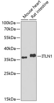
Western blot analysis of various lysates using ITLN1 Rabbit pAb (CAB7234) at 1:1000 dilution. Secondary antibody: HRP-conjugated Goat anti-Rabbit IgG (H+L) (CABS014) at 1:10000 dilution. Lysates/proteins: 25μg per lane. Blocking buffer: 3% nonfat dry milk in TBST. Detection: ECL Basic Kit (AbGn00020). Exposure time: 90s.
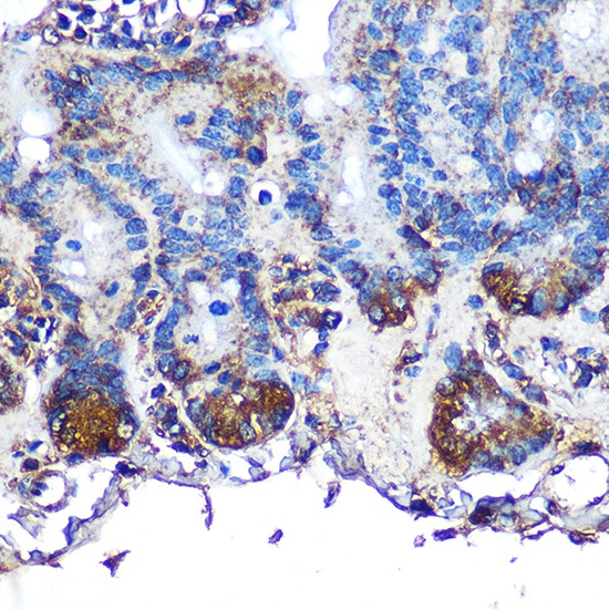
Immunohistochemistry analysis of paraffin-embedded Mouse intestin using ITLN1 Rabbit pAb (CAB7234) at dilution of 1:100 (40x lens). Microwave antigen retrieval performed with 0.01M PBS Buffer (pH 7.2) prior to IHC staining.
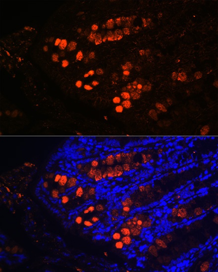
Immunofluorescence analysis of paraffin-embedded mouse colon using ITLN1 Rabbit pAb (CAB7234) at dilution of 1:100 (40x lens). Secondary antibody: Cy3-conjugated Goat anti-Rabbit IgG (H+L) (CABS007) at 1:500 dilution. Blue: DAPI for nuclear staining.