
Description
IL4 Antibody (CAB5649)
The IL4 Antibody (CAB5649) is a high-quality antibody developed for reliable detection and analysis of target proteins. This antibody, produced in rabbits, exhibits high reactivity with human samples and has been validated for use in Western blot applications.IL-4 is known for its role in promoting Th2 cell differentiation, B cell proliferation, and antibody class switching. It is also involved in the regulation of allergic responses and inflammation. The Interleukin-4 Polyclonal Antibody binds specifically to IL-4, allowing for accurate detection and analysis in a variety of cell types.
This antibody is validated for use in WB, IHC-P, ELISA, IF-P applications and has demonstrated reactivity against Human samples.
| Product Name: | IL4 Antibody |
| SKU: | CAB5649 |
| Size: | 20μL, 100μL |
| Reactivity: | Human |
| Conjugate: | Unconjugated |
| Immunogen: | Recombinant protein (or fragment).This information is considered to be commercially sensitive. | ||||||||
| Sequence: | HKCD ITLQ EIIK TLNS LTEQ KTLC TELT VTDI FAAS KNTT EKET FCRA ATVL RQFY SHHE KDTR CLGA TAQQ FHRH KQLI RFLK RLDR NLWG LAGL NSCP VKEA NQST LENF LERL KTIM REKY SKCS S | ||||||||
| Tested Applications: | WB IHC-P ELISA IF-P | ||||||||
| Recommended Dilution: |
| ||||||||
| Synonyms: | BSF1, IL-4, BCGF1, BSF-1, BCGF-1, IL4 |
| Positive Sample: | 293T transfected with IL4 protein |
| Cellular Localization: | Secreted. |
| Calculated MW: | 17kDa |
| Observed MW: | 20kDa |
The protein encoded by this gene is a pleiotropic cytokine produced by activated T cells. This cytokine is a ligand for interleukin 4 receptor. The interleukin 4 receptor also binds to IL13, which may contribute to many overlapping functions of this cytokine and IL13. STAT6, a signal transducer and activator of transcription, has been shown to play a central role in mediating the immune regulatory signal of this cytokine. This gene, IL3, IL5, IL13, and CSF2 form a cytokine gene cluster on chromosome 5q, with this gene particularly close to IL13. This gene, IL13 and IL5 are found to be regulated coordinately by several long-range regulatory elements in an over 120 kilobase range on the chromosome. IL4 is considered an important cytokine for tissue repair, counterbalancing the effects of proinflammatory type 1 cytokines, however, it also promotes allergic airway inflammation. Moreover, IL-4, a type 2 cytokine, mediates and regulates a variety of human host responses such as allergic, anti-parasitic, wound healing, and acute inflammation. This cytokine has been reported to promote resolution of neutrophil-mediated acute lung injury. In an allergic response, IL-4 has an essential role in the production of allergen-specific immunoglobin (Ig) E. This pro-inflammatory cytokine has been observed to be increased in COVID-19 (Coronavirus disease 2019) patients, but is not necessarily associated with severe COVID-19 pathology. Two alternatively spliced transcript variants of this gene encoding distinct isoforms have been reported.
| Purification Method | Affinity purification |
| Gene ID | 3565 |
| RRID | AB_2766409 |
| Buffer Information | Store at -20℃. Avoid freeze / thaw cycles. Buffer: PBS containing 50% glycerol, preserved with proclin300 or sodium azide, pH 7.3. |
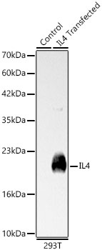 | Western blot analysis of lysates from wild type (WT) and 293T cells transfected with IL4 using IL4 Rabbit pAb (CAB5649) at 1:1000 dilution. Secondary antibody: HRP-conjugated Goat anti-Rabbit IgG (H+L) (CABS014) at 1:10000 dilution. Lysates/proteins: 25 μg per lane. Blocking buffer: 3% nonfat dry milk in TBST. Detection: ECL Basic Kit (AbGn00020). Exposure time: 0.5s. |
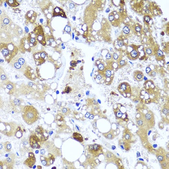 | Immunohistochemistry analysis of paraffin-embedded Human liver damage using IL4 Rabbit pAb (CAB5649) at dilution of 1:100 (40x lens). Microwave antigen retrieval performed with 0.01M PBS Buffer (pH 7.2) prior to IHC staining. |


