The IRAK3 Antibody (CAB5467) is a high-quality antibody developed for reliable detection and analysis of target proteins. This antibody, generated in rabbits, shows high specificity for human samples and has been validated for use in Western blot applications. By binding to the IRAK3 protein, this antibody enables researchers to detect and analyze IRAK3 levels in various cell types, making it ideal for studies in immunology and inflammatory diseases.IRAK3 is a crucial component of the immune system, serving as a negative regulator of inflammatory signaling pathways.
This antibody is validated for use in WB, IF/ICC, ELISA applications and has demonstrated reactivity against Human, Mouse, Rat samples.
Product Name:
IRAK3 Antibody
SKU:
CAB5467
Size:
20μL, 100μL
Reactivity:
Human, Mouse, Rat
Conjugate:
Unconjugated
Immunogen:
Recombinant protein (or fragment).This information is considered to be commercially sensitive.
Recommended starting concentration is 1 μg/mL. Please optimize the concentration based on your specific assay requirements.
Synonyms:
ASRT5, IRAKM, IRAK3
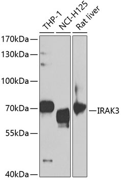
Positive Sample:
THP-1, NCI-H125, Rat liver
Cellular Localization:
Cytoplasm, Nucleus, Plasma Membrane.
Calculated MW:
68kDa
Observed MW:
68kDa
This gene encodes a member of the interleukin-1 receptor-associated kinase protein family. Members of this family are essential components of the Toll/IL-R immune signal transduction pathways. This protein is primarily expressed in monocytes and macrophages and functions as a negative regulator of Toll-like receptor signaling. Mutations in this gene are associated with a susceptibility to asthma. Alternate splicing results in multiple transcript variants.
Purification Method
Affinity purification
Gene ID
11213
RRID
AB_2766268
Buffer Information
Store at -20℃. Avoid freeze / thaw cycles. Buffer: PBS containing 50% glycerol, preserved with proclin300 or sodium azide, pH 7.3.
Western blot analysis of various lysates using IRAK3 Rabbit pAb (CAB5467) at 1:1000 dilution. Secondary antibody: HRP-conjugated Goat anti-Rabbit IgG (H+L) (CABS014) at 1:10000 dilution. Lysates/proteins: 25μg per lane. Blocking buffer: 3% nonfat dry milk in TBST. Detection: ECL Basic Kit (AbGn00020). Exposure time: 90s.
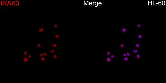
Immunofluorescence analysis of HL-60 cells using IRAK3 Rabbit pAb(CAB5467) at a dilution of 1:100 (40x lens). Secondary antibody:Cy3 Goat Anti-Rabbit IgG (H+L)(CABS007) at 1:500 dilution. Blue: DAPI for nuclear staining.