The JAG2 Antibody (CAB14247) is a high-quality antibody developed for reliable detection and analysis of target proteins. This antibody, generated using rabbits, shows high reactivity towards human samples and is validated for use in Western blot applications, allowing for the detection and analysis of JAG2 in various cell types. Its specificity and efficiency make it an essential component in studies related to developmental biology, cancer research, and regenerative medicine.JAG2 is a transmembrane protein known for its interactions with Notch receptors, leading to the activation of downstream signaling cascades that control cell differentiation, proliferation, and survival.
This antibody is validated for use in WB, IF/ICC, ELISA applications and has demonstrated reactivity against Human, Mouse, Rat samples.
Product Name:
JAG2 Antibody
SKU:
CAB14247
Size:
20μL, 100μL
Reactivity:
Human, Mouse, Rat
Conjugate:
Unconjugated
Immunogen:
Recombinant protein (or fragment).This information is considered to be commercially sensitive.
Recommended starting concentration is 1 μg/mL. Please optimize the concentration based on your specific assay requirements.
Synonyms:
HJ2, SER2, LGMDR27, JAG2
Positive Sample:
Mouse heart
Cellular Localization:
Membrane, Single-Pass Type I Membrane Protein.
Calculated MW:
133kDa
Observed MW:
150kDa
The Notch signaling pathway is an intercellular signaling mechanism that is essential for proper embryonic development. Members of the Notch gene family encode transmembrane receptors that are critical for various cell fate decisions. The protein encoded by this gene is one of several ligands that activate Notch and related receptors. Two transcript variants encoding different isoforms have been found for this gene.
Purification Method
Affinity purification
Gene ID
3714
RRID
AB_2761107
Buffer Information
Store at -20℃. Avoid freeze / thaw cycles. Buffer: PBS with 0.01% thimerosal,50% glycerol,pH7.3.
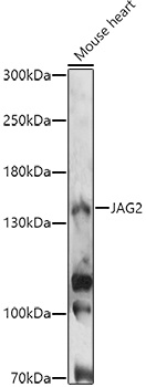
Western blot analysis of lysates from Mouse heart, using JAG2 Rabbit pAb (CAB14247) at 1:1000 dilution. Secondary antibody: HRP-conjugated Goat anti-Rabbit IgG (H+L) (CABS014) at 1:10000 dilution. Lysates/proteins: 25μg per lane. Blocking buffer: 3% nonfat dry milk in TBST. Detection: ECL Enhanced Kit (AbGn00021). Exposure time: 20s.
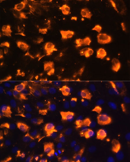
Immunofluorescence analysis of C6 cells using JAG2 Rabbit pAb (CAB14247) at dilution of 1:100. Blue: DAPI for nuclear staining.