The JPH1 Antibody (CAB18476) is a high-quality antibody developed for reliable detection and analysis of target proteins. Raised in rabbits, this antibody is highly specific and reactive with human samples, making it suitable for use in Western blot applications. By binding to JPH1, researchers can detect and analyze this protein in various cell types, making it an ideal tool for studies in cell biology and neuroscience.JPH1 is known for its role in organizing membrane structures in cells, specifically in the junctions between the plasma membrane and the endoplasmic/sarcoplasmic reticulum. This unique function makes JPH1 a key player in maintaining cellular structure and communication.
This antibody is validated for use in WB, ELISA applications and has demonstrated reactivity against Mouse samples.
Product Name:
JPH1 Antibody
SKU:
CAB18476
Size:
20μL, 100μL
Reactivity:
Mouse
Immunogen:
Recombinant protein (or fragment).This information is considered to be commercially sensitive.
Recommended starting concentration is 1 μg/mL. Please optimize the concentration based on your specific assay requirements.
Synonyms:
JP1, JP-1, CMT2K, JPH1
Positive Sample:
Mouse skeletal muscle
Cellular Localization:
Junctional Membrane Complex, Nucleoplasm, Plasma Membrane, Sarcoplasmic Reticulum, Z Disc.
Calculated MW:
72kDa
Observed MW:
62kDa
Junctional complexes between the plasma membrane and endoplasmic/sarcoplasmic reticulum are a common feature of all excitable cell types and mediate cross talk between cell surface and intracellular ion channels. The protein encoded by this gene is a component of junctional complexes and is composed of a C-terminal hydrophobic segment spanning the endoplasmic/sarcoplasmic reticulum membrane and a remaining cytoplasmic domain that shows specific affinity for the plasma membrane. This gene is a member of the junctophilin gene family.
Purification Method
Affinity purification
Gene ID
56704
RRID
AB_2862243
Buffer Information
Store at -20℃. Avoid freeze / thaw cycles. Buffer: PBS with 0.01% thimerosal,50% glycerol,pH7.3.
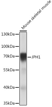
Western blot analysis of lysates from Mouse skeletal muscle, using JPH1 Rabbit pAb (CAB18476) at 1:1000 dilution. Secondary antibody: HRP-conjugated Goat anti-Rabbit IgG (H+L) (CABS014) at 1:10000 dilution. Lysates/proteins: 25μg per lane. Blocking buffer: 3% nonfat dry milk in TBST. Detection: ECL Basic Kit (AbGn00020). Exposure time: 1s.