The KLK1 Antibody (CAB1807) is a high-quality antibody developed for reliable detection and analysis of target proteins. This antibody, produced in rabbits, is highly specific to human samples and has been validated for use in Western blot applications.Kallikrein 1, also known as tissue kallikrein, is known to be involved in the modulation of inflammatory responses and is overexpressed in certain cancers, making it a target of interest in cancer research. The Kallikrein 1 Polyclonal Antibody enables detection and analysis of Kallikrein 1 protein levels in different cell types, allowing researchers to further explore its role in disease pathology and potential therapeutic applications.
This antibody is validated for use in WB, IHC-P, ELISA applications and has demonstrated reactivity against Human, Mouse, Rat samples.
Product Name:
KLK1 Antibody
SKU:
CAB1807
Size:
20μL, 100μL
Reactivity:
Human, Mouse, Rat
Conjugate:
Unconjugated
Immunogen:
Recombinant protein (or fragment).This information is considered to be commercially sensitive.
Recommended starting concentration is 1 μg/mL. Please optimize the concentration based on your specific assay requirements.
Synonyms:
hK1, KLKR, Klk6, KLK1
Positive Sample:
SW480, K-562
Cellular Localization:
Extracellular Exosome, Nucleus.
Calculated MW:
29kDa
Observed MW:
22kDa
Kallikreins are a subgroup of serine proteases having diverse physiological functions. Growing evidence suggests that many kallikreins are implicated in carcinogenesis and some have potential as novel cancer and other disease biomarkers. This gene is one of the fifteen kallikrein subfamily members located in a cluster on chromosome 19. This protein is functionally conserved in its capacity to release the vasoactive peptide, Lys-bradykinin, from low molecular weight kininogen.
Purification Method
Affinity purification
Gene ID
3816
RRID
AB_2763846
Buffer Information
Store at -20℃. Avoid freeze / thaw cycles. Buffer: PBS containing 50% glycerol, preserved with proclin300 or sodium azide, pH 7.3.
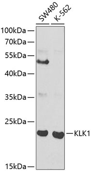
Western blot analysis of various lysates using KLK1 Rabbit pAb (CAB1807) at 1:1000 dilution. Secondary antibody: HRP-conjugated Goat anti-Rabbit IgG (H+L) (CABS014) at 1:10000 dilution. Lysates/proteins: 25μg per lane. Blocking buffer: 3% nonfat dry milk in TBST.
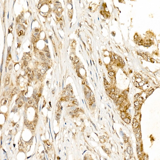
Immunohistochemistry analysis of paraffin-embedded Human colon carcinoma using KLK1 Rabbit pAb (CAB1807) at dilution of 1:500 (40x lens). High pressure antigen retrieval performed with 0.01M Citrate buffer (pH 6.0) prior to IHC staining.
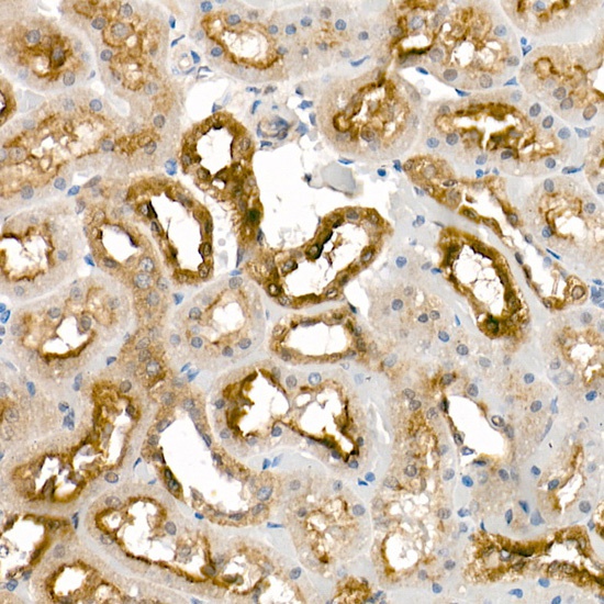
Immunohistochemistry analysis of paraffin-embedded Rat kidney using KLK1 Rabbit pAb (CAB1807) at dilution of 1:500 (40x lens). High pressure antigen retrieval performed with 0.01M Citrate buffer (pH 6.0) prior to IHC staining.