The KLK6 Antibody (CAB12055) is a high-quality antibody developed for reliable detection and analysis of target proteins. This antibody, generated from rabbits, is highly specific for detecting Kallikrein-6 in human samples and has been validated for use in Western blot applications.Kallikrein-6, also known as Neurosin, is involved in the proteolytic degradation of proteins and peptides, playing a role in processes such as neurodegeneration, cancer progression, and inflammation. Its expression and activity have been linked to conditions such as Alzheimer's disease, ovarian cancer, and multiple sclerosis, making it a promising target for diagnostic and therapeutic interventions.
This antibody is validated for use in WB, ELISA applications and has demonstrated reactivity against Human samples.
Product Name:
KLK6 Antibody
SKU:
CAB12055
Size:
20μL, 100μL
Reactivity:
Human
Conjugate:
Unconjugated
Immunogen:
Synthetic peptide. This information is considered to be commercially sensitive.
This gene encodes a member of the kallikrein subfamily of the peptidase S1 family of serine proteases. Growing evidence suggests that many kallikreins are implicated in carcinogenesis and some have potential as novel cancer and other disease biomarkers. The encoded preproprotein is proteolytically processed to generate the mature protease. Expression of this protease is regulated by steroid hormones and may be elevated in multiple human cancers and in serum from psoriasis patients. The encoded protease may participate in the cleavage of amyloid precursor protein and alpha-synuclein, thus implicating this protease in Alzheimer's and Parkinson's disease, respectively. This gene is located in a gene cluster on chromosome 19. Alternative splicing results in multiple transcript variants, at least one of which encodes an isoform that is proteolytically processed.
Purification Method
Affinity purification
Gene ID
5653
RRID
AB_2758964
Buffer Information
Store at -20℃. Avoid freeze / thaw cycles. Buffer: PBS with 0.01% thimerosal,50% glycerol,pH7.3.
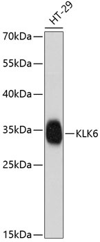
Western blot analysis of lysates from HT-29 cells, using KLK6 Rabbit pAb (CAB12055) at 1:1000 dilution. Secondary antibody: HRP-conjugated Goat anti-Rabbit IgG (H+L) (CABS014) at 1:10000 dilution. Lysates/proteins: 25μg per lane. Blocking buffer: 3% nonfat dry milk in TBST. Detection: ECL Enhanced Kit (AbGn00021). Exposure time: 90s.