The KDM7A Antibody (CAB14692) is a high-quality antibody developed for reliable detection and analysis of target proteins. This antibody, produced in rabbits, exhibits high reactivity with human samples and has been validated for use in Western blot and immunohistochemistry applications.KDM7A is known to play a key role in gene expression regulation through histone modification processes, making it a key player in cellular processes such as differentiation, development, and DNA damage response. The KDM7A Polyclonal Antibody binds specifically to the KDM7A protein, enabling precise detection and analysis in various cell types.
This antibody is validated for use in WB, ELISA applications and has demonstrated reactivity against Human samples.
Product Name:
KDM7A Antibody
SKU:
CAB14692
Size:
20μL, 100μL
Reactivity:
Human
Conjugate:
Unconjugated
Immunogen:
Recombinant protein (or fragment).This information is considered to be commercially sensitive.
Recommended starting concentration is 1 μg/mL. Please optimize the concentration based on your specific assay requirements.
Synonyms:
Jhdm1d, mKIAA1718, A630082K20Rik, KDM7A
Positive Sample:
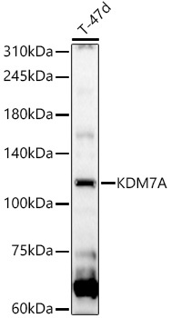
T-47d
Cellular Localization:
Nucleus.
Calculated MW:
106kDa
Observed MW:
106kDa
Enables histone H3-methyl-lysine-9 demethylase activity and histone H3-tri/di-methyl-lysine-27 demethylase activity. Involved in histone H3-K27 demethylation; histone H3-K9 demethylation; and positive regulation of transcription, DNA-templated. Predicted to be located in nucleolus and nucleoplasm. Human ortholog(s) of this gene implicated in melanoma. Orthologous to human KDM7A (lysine demethylase 7A).
Purification Method
Affinity purification
Gene ID
338523
RRID
AB_2761567
Buffer Information
Store at -20℃. Avoid freeze / thaw cycles. Buffer: PBS containing 50% glycerol, preserved with proclin300 or sodium azide, pH 7.3.
Western blot analysis of lysates from T-47d cells, using KDM7A Rabbit pAb (CAB14692) at 1:1000 dilution. Secondary antibody: HRP-conjugated Goat anti-Rabbit IgG (H+L) (CABS014) at 1:10000 dilution. Lysates/proteins: 25μg per lane. Blocking buffer: 3% nonfat dry milk in TBST. Detection: ECL Basic Kit (AbGn00020). Exposure time: 30s.