The KIF5A Antibody (CAB3303) is a high-quality antibody developed for reliable detection and analysis of target proteins. This antibody, generated in rabbits, exhibits high reactivity with human samples and has been rigorously validated for Western blotting applications. By specifically binding to the KIF5A protein, this antibody enables precise detection and analysis in a variety of cell types, making it an essential component for studies in cell biology and neuroscience.
This antibody is validated for use in WB, IF/ICC, ELISA applications and has demonstrated reactivity against Human, Mouse, Rat samples.
Product Name:
KIF5A Antibody
SKU:
CAB3303
Size:
20μL, 100μL
Reactivity:
Human, Mouse, Rat
Conjugate:
Unconjugated
Immunogen:
Recombinant protein (or fragment).This information is considered to be commercially sensitive.
Recommended starting concentration is 1 μg/mL. Please optimize the concentration based on your specific assay requirements.
Synonyms:
NKHC, ALS25, MY050, NEIMY, SPG10, D12S1889, KIF5A
Positive Sample:
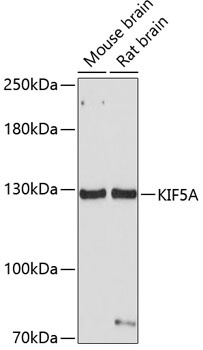
Mouse brain, Rat brain
Cellular Localization:
Cytoplasm, Cytoskeleton, Perinuclear Region.
Calculated MW:
117kDa
Observed MW:
129kDa
This gene encodes a member of the kinesin family of proteins. Members of this family are part of a multisubunit complex that functions as a microtubule motor in intracellular organelle transport. Mutations in this gene cause autosomal dominant spastic paraplegia 10.
Purification Method
Affinity purification
Gene ID
3798
RRID
AB_2765035
Buffer Information
Store at -20℃. Avoid freeze / thaw cycles. Buffer: PBS containing 50% glycerol, preserved with proclin300 or sodium azide, pH 7.3.
Western blot analysis of various lysates using KIF5A Rabbit pAb (CAB3303) at 1:1000 dilution. Secondary antibody: HRP-conjugated Goat anti-Rabbit IgG (H+L) (CABS014) at 1:10000 dilution. Lysates/proteins: 25μg per lane. Blocking buffer: 3% nonfat dry milk in TBST. Detection: ECL Basic Kit (AbGn00020). Exposure time: 30s.
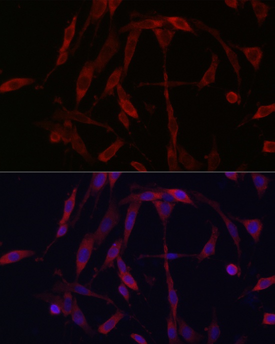
Immunofluorescence analysis of PC-12 cells using KIF5A Rabbit pAb (CAB3303) at dilution of 1:200 (40x lens). Secondary antibody: Cy3-conjugated Goat anti-Rabbit IgG (H+L) (CABS007) at 1:500 dilution. Blue: DAPI for nuclear staining.