The KIR3DL1 Antibody (CAB1617) is a high-quality antibody developed for reliable detection and analysis of target proteins. This antibody, produced in rabbits, exhibits high reactivity with human samples and is validated for use in Western blot applications. By specifically binding to the KIR3DL1 protein, this antibody enables precise detection and analysis in a variety of cell types, making it an indispensable asset for investigations in immunology and cancer research.
This antibody is validated for use in WB, ELISA applications and has demonstrated reactivity against Human, Mouse samples.
Product Name:
KIR3DL1 Antibody
SKU:
CAB1617
Size:
20μL, 100μL
Reactivity:
Human, Mouse
Conjugate:
Unconjugated
Immunogen:
Recombinant protein (or fragment).This information is considered to be commercially sensitive.
Recommended starting concentration is 1 μg/mL. Please optimize the concentration based on your specific assay requirements.
Synonyms:
KIR, NKB1, NKAT3, NKB1B, NKAT-3, CD158E1, KIR2DL5B, KIR3DL1/S1, KIR3DL1
Positive Sample:
DU145, BT-474, U-937, Raji, Mouse thymus
Cellular Localization:
Cell Membrane, Single-Pass Type I Membrane Protein.
Calculated MW:
49kDa
Observed MW:
48kDa
Killer cell immunoglobulin-like receptors (KIRs) are transmembrane glycoproteins expressed by natural killer cells and subsets of T cells. The KIR genes are polymorphic and highly homologous and they are found in a cluster on chromosome 19q13.4 within the 1 Mb leukocyte receptor complex (LRC). The gene content of the KIR gene cluster varies among haplotypes, although several "framework" genes are found in all haplotypes (KIR3DL3, KIR3DP1, KIR3DL4, KIR3DL2). The KIR proteins are classified by the number of extracellular immunoglobulin domains (2D or 3D) and by whether they have a long (L) or short (S) cytoplasmic domain. KIR proteins with the long cytoplasmic domain transduce inhibitory signals upon ligand binding via an immune tyrosine-based inhibitory motif (ITIM), while KIR proteins with the short cytoplasmic domain lack the ITIM motif and instead associate with the TYRO protein tyrosine kinase binding protein to transduce activating signals. The ligands for several KIR proteins are subsets of HLA class I molecules; thus, KIR proteins are thought to play an important role in regulation of the immune response.
Purification Method
Affinity purification
Gene ID
3811
RRID
AB_2763619
Buffer Information
Store at -20℃. Avoid freeze / thaw cycles. Buffer: PBS containing 50% glycerol, preserved with proclin300 or sodium azide, pH 7.3.
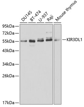
Western blot analysis of various lysates using KIR3DL1 Rabbit pAb (CAB1617) at 1:1000 dilution. Secondary antibody: HRP-conjugated Goat anti-Rabbit IgG (H+L) (CABS014) at 1:10000 dilution. Lysates/proteins: 25μg per lane. Blocking buffer: 3% nonfat dry milk in TBST.
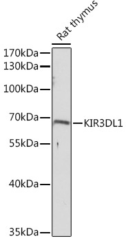
Western blot analysis of lysates from Rat thymus, using KIR3DL1 Rabbit pAb (CAB1617) at 1:3000 dilution. Secondary antibody: HRP-conjugated Goat anti-Rabbit IgG (H+L) (CABS014) at 1:10000 dilution. Lysates/proteins: 25μg per lane. Blocking buffer: 3% nonfat dry milk in TBST. Detection: ECL Basic Kit (AbGn00020). Exposure time: 90s.