The KLRD1 Antibody (CAB12698) is a high-quality antibody developed for reliable detection and analysis of target proteins. This antibody, produced in rabbits, exhibits high reactivity with human samples and is validated for use in Western blot applications. By specifically binding to the KLRD1 protein, this antibody enables accurate detection and analysis in a variety of cell types, making it an invaluable asset for studies in immunology and cancer research.KLRD1, a member of the C-type lectin-like receptor family, plays a crucial role in immune surveillance and response. It is involved in the recognition of HLA-E molecules, which are essential for the regulation of immune responses to infected or stressed cells.
This antibody is validated for use in WB, ELISA applications and has demonstrated reactivity against Human, Mouse, Rat samples.
Product Name:
KLRD1 Antibody
SKU:
CAB12698
Size:
20μL, 100μL
Reactivity:
Human, Mouse, Rat
Conjugate:
Unconjugated
Immunogen:
Recombinant protein (or fragment).This information is considered to be commercially sensitive.
Recommended starting concentration is 1 μg/mL. Please optimize the concentration based on your specific assay requirements.
Synonyms:
CD94, KLRD1
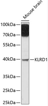
Positive Sample:
Mouse brain
Cellular Localization:
Membrane, Single-Pass Type Ii Membrane Protein.
Calculated MW:
21kDa
Observed MW:
40kDa
Natural killer (NK) cells are a distinct lineage of lymphocytes that mediate cytotoxic activity and secrete cytokines upon immune stimulation. Several genes of the C-type lectin superfamily, including members of the NKG2 family, are expressed by NK cells and may be involved in the regulation of NK cell function. KLRD1 (CD94) is an antigen preferentially expressed on NK cells and is classified as a type II membrane protein because it has an external C terminus. Several transcript variants encoding different isoforms have been found for this gene.
Purification Method
Affinity purification
Gene ID
3824
RRID
AB_2759541
Buffer Information
Store at -20℃. Avoid freeze / thaw cycles. Buffer: PBS with 0.01% thimerosal,50% glycerol,pH7.3.
Western blot analysis of lysates from mouse brain, using KLRD1 Rabbit pAb (CAB12698) at 1:3000 dilution. Secondary antibody: HRP-conjugated Goat anti-Rabbit IgG (H+L) (CABS014) at 1:10000 dilution. Lysates/proteins: 25μg per lane. Blocking buffer: 3% nonfat dry milk in TBST. Detection: ECL Enhanced Kit (AbGn00021). Exposure time: 90s.