The [KO Validated] AKT2 Antibody (CAB18019) is a high-quality antibody developed for reliable detection and analysis of target proteins. This antibody, produced in rabbits, is highly specific for human samples and has been validated for use in Western blot applications.AKT2, a member of the AKT family of serine/threonine protein kinases, is known for its role in various cellular processes, including cell proliferation, differentiation, and metabolism. Dysregulation of AKT2 signaling is commonly associated with cancer and other diseases, making it a promising target for therapeutic interventions.
This antibody is validated for use in WB, ELISA applications and has demonstrated reactivity against Human, Mouse samples.
Product Name:
[KO Validated] AKT2 Antibody
SKU:
CAB18019
Size:
20μL, 100μL
Reactivity:
Human, Mouse
Conjugate:
Unconjugated
Immunogen:
Synthetic peptide. This information is considered to be commercially sensitive.
Recommended starting concentration is 1 μg/mL. Please optimize the concentration based on your specific assay requirements.
Synonyms:
PKBB, PRKBB, HIHGHH, PKBBETA, RAC-BETA, AKT2
Positive Sample:
293T, Raji, Jurkat, HeLa, LO2, Mouse kidney
Cellular Localization:
Cell Membrane, Cytoplasm, Early Endosome, Nucleus, Peripheral Membrane Protein.
Calculated MW:
56kDa
Observed MW:
60kDa
This gene is a putative oncogene encoding a protein belonging to a subfamily of serine/threonine kinases containing SH2-like (Src homology 2-like) domains, which is involved in signaling pathways. The gene serves as an oncogene in the tumorigenesis of cancer cells For example, its overexpression contributes to the malignant phenotype of a subset of human ductal pancreatic cancers. The encoded protein is a general protein kinase capable of phophorylating several known proteins, and has also been implicated in insulin signaling.
Purification Method
Affinity purification
Gene ID
208
RRID
AB_2861815
Buffer Information
Store at -20℃. Avoid freeze / thaw cycles. Buffer: PBS with 0.01% thimerosal,50% glycerol,pH7.3.
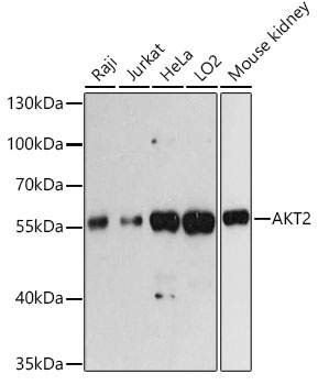
Western blot analysis of various lysates using AKT2 Rabbit pAb (CAB18019) at 1:3000 dilution. Secondary antibody: HRP-conjugated Goat anti-Rabbit IgG (H+L) (CABS014) at 1:10000 dilution. Lysates/proteins: 25μg per lane. Blocking buffer: 3% nonfat dry milk in TBST. Detection: ECL Basic Kit (AbGn00020). Exposure time: 60s.
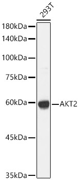
Western blot analysis of lysates from 293T cells, using AKT2 Rabbit pAb (CAB18019) at 1:1000 dilution. Secondary antibody: HRP-conjugated Goat anti-Rabbit IgG (H+L) (CABS014) at 1:10000 dilution. Lysates/proteins: 25μg per lane. Blocking buffer: 3% nonfat dry milk in TBST. Detection: ECL Basic Kit (AbGn00020). Exposure time: 1s.