The [KO Validated] GNAI3 Antibody (CAB13307) is a high-quality antibody developed for reliable detection and analysis of target proteins. This antibody, produced in rabbits, demonstrates high specificity and sensitivity towards human samples, making it a reliable tool for Western blot applications. By binding specifically to GNAI3, this antibody enables the detection and analysis of the protein in various cell types, making it an essential component for studies in cell signaling and cancer research.GNAI3, a member of the G protein family, plays a crucial role in mediating intracellular signaling cascades triggered by various extracellular stimuli.
This antibody is validated for use in WB, IF/ICC, IP, ELISA applications and has demonstrated reactivity against Human, Mouse samples.
Product Name:
[KO Validated] GNAI3 Antibody
SKU:
CAB13307
Size:
20μL, 100μL
Reactivity:
Human, Mouse
Conjugate:
Unconjugated
Immunogen:
Recombinant protein (or fragment).This information is considered to be commercially sensitive.
Guanine nucleotide-binding proteins (G proteins) are involved as modulators or transducers in various transmembrane signaling pathways. G proteins are composed of 3 units: alpha, beta and gamma. This gene encodes an alpha subunit and belongs to the G-alpha family. Mutation in this gene, resulting in a gly40-to-arg substitution, is associated with auriculocondylar syndrome, and shown to affect downstream targets in the G protein-coupled endothelin receptor pathway.
Purification Method
Affinity purification
Gene ID
2773
RRID
AB_2861683
Buffer Information
Store at -20℃. Avoid freeze / thaw cycles. Buffer: PBS containing 50% glycerol, preserved with proclin300 or sodium azide, pH 7.3.
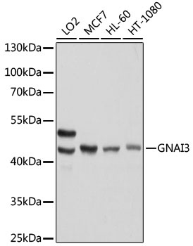
Western blot analysis of various lysates using [KO Validated] GNAI3 Rabbit pAb (CAB13307) at 1:1000 dilution. Secondary antibody: HRP-conjugated Goat anti-Rabbit IgG (H+L) (CABS014) at 1:10000 dilution. Lysates/proteins: 25μg per lane. Blocking buffer: 3% nonfat dry milk in TBST. Detection: ECL Basic Kit (AbGn00020). Exposure time: 1s.
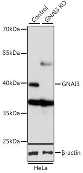
Western blot analysis of lysates from wild type (WT) and GNAI3 knockout (KO) HeLa cells, using [KO Validated] GNAI3 Rabbit pAb (CAB13307) at 1:1000 dilution. Secondary antibody: HRP-conjugated Goat anti-Rabbit IgG (H+L) (CABS014) at 1:10000 dilution. Lysates/proteins: 25μg per lane. Blocking buffer: 3% nonfat dry milk in TBST. Detection: ECL Basic Kit (AbGn00020). Exposure time: 1s.
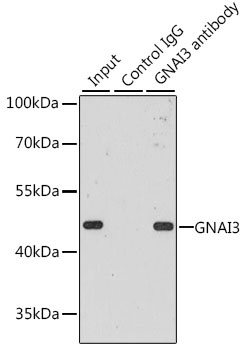
Immunoprecipitation analysis of 200 μg extracts of MCF-7 cells, using 3 μg GNAI3 antibody (CAB13307). Western blot was performed from the immunoprecipitate using GNAI3 antibody (CAB13307) at a dilution of 1:1000.