The GM130 Monoclonal Antibody (CAB11408) is a high-quality antibody developed for reliable detection and analysis of target proteins. This monoclonal antibody, produced in rabbits, is optimized for use in various applications such as immunofluorescence, immunohistochemistry, and Western blotting.GM130 is essential for proper Golgi organization and vesicle trafficking, playing a crucial role in intracellular transport and protein glycosylation. Dysregulation of GM130 has been implicated in various human diseases, including cancer and neurodegenerative disorders.
This antibody is validated for use in WB, IHC-P, IF/ICC, ELISA applications and has demonstrated reactivity against Human, Mouse samples.
Product Name:
GM130 Monoclonal Antibody
SKU:
CAB11408
Size:
20μL, 100μL
Reactivity:
Human, Mouse
Clone Number:
ARC0589
Conjugate:
Unconjugated
Immunogen:
Synthetic peptide. This information is considered to be commercially sensitive.
The Golgi apparatus, which participates in glycosylation and transport of proteins and lipids in the secretory pathway, consists of a series of stacked cisternae (flattened membrane sacs). Interactions between the Golgi and microtubules are thought to be important for the reorganization of the Golgi after it fragments during mitosis. This gene encodes one of the golgins, a family of proteins localized to the Golgi. This encoded protein has been postulated to play roles in the stacking of Golgi cisternae and in vesicular transport. Several alternatively spliced transcript variants of this gene have been described, but the full-length nature of these variants has not been determined.
Purification Method
Affinity purification
Gene ID
2801
RRID
AB_2861561
Buffer Information
Store at -20℃. Avoid freeze / thaw cycles. Buffer: PBS containing 50% glycerol and 0.05% BSA, preserved with proclin300 or sodium azide, pH 7.3.
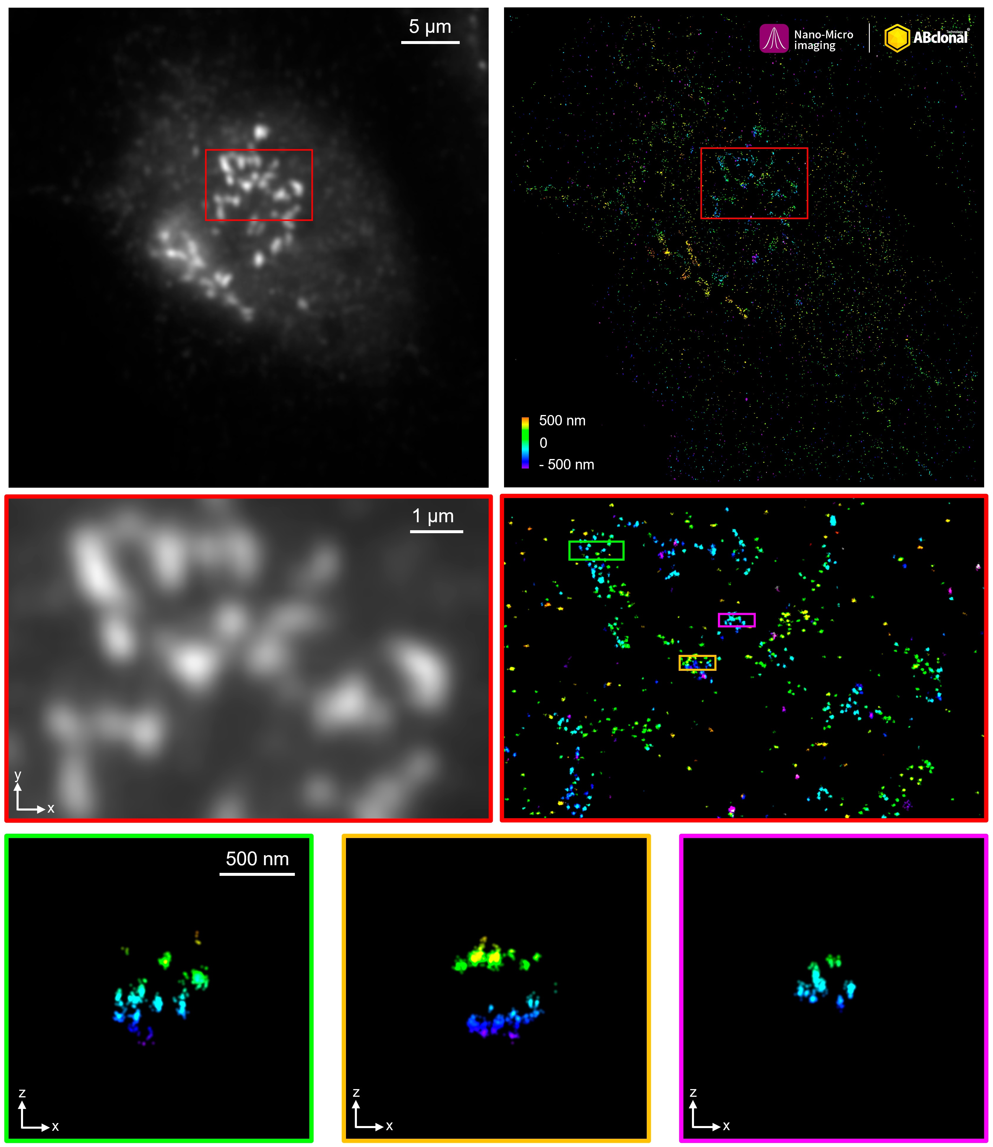
The STORM super-resolution (SR) imaging of U-2 OS cells using GM130 Rabbit mAb (CAB11408, ABclonal) at dilution of 1:200 with 3% paraformaldehyde (PFA) +0.1% glutaraldehyde (GA) fixation. The immunostaining was performed by Full Automatic Immunofluorescence Workflow System (Workflow Ultra300, Nano-Micro imaging, China). Image was performed with Single-Molecule Localization Super-Resolution Microscopy (STORM Ultra300, Nano-Micro imaging, China). We acknowledge Nano-Micro imaging Biotechnology Co., Ltd. in SR image processing and kindly providing this image.
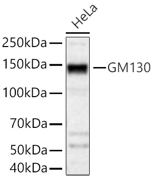
Western blot analysis of lysates from HeLa cells using GM130 Rabbit mAb (CAB11408) at 1:1000 dilution incubated overnight at 4℃. Secondary antibody: HRP-conjugated Goat anti-Rabbit IgG (H+L) (CABS014) at 1:10000 dilution. Lysates/proteins: 25 μg per lane. Blocking buffer: 3% nonfat dry milk in TBST. Detection: ECL Basic Kit (AbGn00020). Exposure time: 10s.
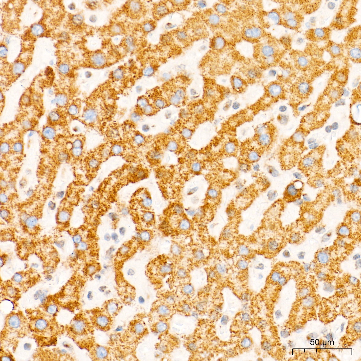
Immunohistochemistry analysis of paraffin-embedded Human liver tissue using GM130 Rabbit mAb (CAB11408) at a dilution of 1:200 (40x lens). High pressure antigen retrieval performed with 0.01M Tris-EDTA Buffer (pH 9.0) prior to IHC staining.
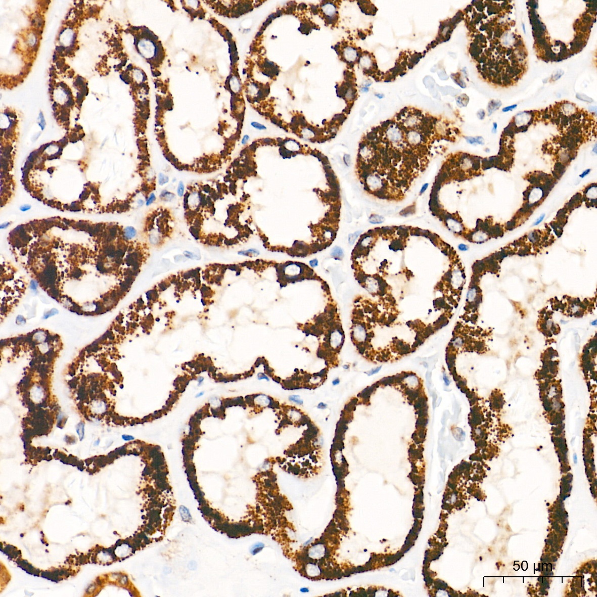
Immunohistochemistry analysis of paraffin-embedded Human kidney tissue using GM130 Rabbit mAb (CAB11408) at a dilution of 1:200 (40x lens). High pressure antigen retrieval performed with 0.01M Tris-EDTA Buffer (pH 9.0) prior to IHC staining.