The [KO Validated] MEK2 Antibody (CAB14770) is a high-quality antibody developed for reliable detection and analysis of target proteins. This antibody, produced in rabbits, exhibits high specificity and sensitivity towards human samples and has been rigorously validated for use in various experimental applications.MAP2K2, also known as MEK2, plays a crucial role in cell proliferation, differentiation, and survival by transmitting extracellular signals to the nucleus. Dysregulation of the MAPK pathway, in which MAP2K2 is a critical component, has been implicated in various diseases including cancer, neurodegenerative disorders, and cardiovascular diseases.
This antibody is validated for use in WB, IHC-P, IF/ICC, ELISA applications and has demonstrated reactivity against Human, Mouse, Rat samples.
Product Name:
[KO Validated] MEK2 Antibody
SKU:
CAB14770
Size:
20μL, 100μL
Reactivity:
Human, Mouse, Rat
Conjugate:
Unconjugated
Immunogen:
Synthetic peptide. This information is considered to be commercially sensitive.
Recommended starting concentration is 1 μg/mL. Please optimize the concentration based on your specific assay requirements.
Synonyms:
CFC4, MEK2, MKK2, MAPKK2, PRKMK2, K2
Positive Sample:
HeLa, 293T, HT-29
Cellular Localization:
Cytoplasm, Membrane, Peripheral Membrane Protein.
Calculated MW:
44kDa
Observed MW:
45kDa
The protein encoded by this gene is a dual specificity protein kinase that belongs to the MAP kinase kinase family. This kinase is known to play a critical role in mitogen growth factor signal transduction. It phosphorylates and thus activates MAPK1/ERK2 and MAPK2/ERK3. The activation of this kinase itself is dependent on the Ser/Thr phosphorylation by MAP kinase kinase kinases. Mutations in this gene cause cardiofaciocutaneous syndrome (CFC syndrome), a disease characterized by heart defects, cognitive disability, and distinctive facial features similar to those found in Noonan syndrome. The inhibition or degradation of this kinase is also found to be involved in the pathogenesis of Yersinia and anthrax. A pseudogene, which is located on chromosome 7, has been identified for this gene.
Purification Method
Affinity purification
Gene ID
5605
RRID
AB_2861706
Buffer Information
Store at -20℃. Avoid freeze / thaw cycles. Buffer: PBS with 0.01% thimerosal,50% glycerol,pH7.3.
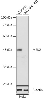
Western blot analysis of lysates from wild type (WT) and MEK2 knockout (KO) HeLa cells, using [KO Validated] MEK2 Rabbit pAb (CAB14770) at 1:1000 dilution. Secondary antibody: HRP-conjugated Goat anti-Rabbit IgG (H+L) (CABS014) at 1:10000 dilution. Lysates/proteins: 25μg per lane. Blocking buffer: 3% nonfat dry milk in TBST. Detection: ECL Basic Kit (AbGn00020). Exposure time: 90s.
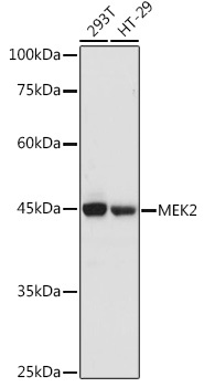
Western blot analysis of various lysates using [KO Validated] MEK2 Rabbit pAb (CAB14770) at 1:1000 dilution. Secondary antibody: HRP-conjugated Goat anti-Rabbit IgG (H+L) (CABS014) at 1:10000 dilution. Lysates/proteins: 25μg per lane. Blocking buffer: 3% nonfat dry milk in TBST. Detection: ECL Basic Kit (AbGn00020). Exposure time: 90s.
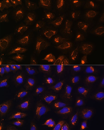
Immunofluorescence analysis of U2OS cells using [KO Validated] MEK2 Rabbit pAb (CAB14770) at dilution of 1:100. Blue: DAPI for nuclear staining.