The [KO Validated] MAP3K1 Antibody (CAB18041) is a high-quality antibody developed for reliable detection and analysis of target proteins. The protein encoded by this gene is a serine/threonine kinase and is part of some signal transduction cascades, including the ERK and JNK kinase pathways as well as the NF-kappa-B pathway. The encoded protein is activated by autophosphorylation and requires magnesium as a cofactor in phosphorylating other proteins. This protein has E3 ligase activity conferred by a plant homeodomain (PHD) in its N-terminus and phospho-kinase activity conferred by a kinase domain in its C-terminus.
This antibody is validated for use in WB, IHC-P, ELISA applications and has demonstrated reactivity against Human, Mouse, Rat samples.
Product Name:
[KO Validated] MAP3K1 Antibody
SKU:
CAB18041
Size:
100μL, 20μL
Reactivity:
Human, Mouse, Rat
Conjugate:
Unconjugated
Immunogen:
Synthetic peptide. This information is considered to be commercially sensitive.
Tested Applications:
WBIHC-PELISA
Recommended Dilution:
WB
1:500 - 1:2000
IHC-P
1:50 - 1:200
ELISA
Recommended starting concentration is 1 μg/mL. Please optimize the concentration based on your specific assay requirements.
Synonyms:
MEKK, MEKK1, SRXY6, MEKK 1, MAPKKK1, K1
Positive Sample:
HeLa, Raji, A-431
Cellular Localization:
Cytoplasm, Cytosol.
Calculated MW:
164kDa
Observed MW:
164kDa
The protein encoded by this gene is a serine/threonine kinase and is part of some signal transduction cascades, including the ERK and JNK kinase pathways as well as the NF-kappa-B pathway. The encoded protein is activated by autophosphorylation and requires magnesium as a cofactor in phosphorylating other proteins. This protein has E3 ligase activity conferred by a plant homeodomain (PHD) in its N-terminus and phospho-kinase activity conferred by a kinase domain in its C-terminus.
Purification Method
Affinity purification
Gene ID
4214
RRID
AB_2861837
Buffer Information
Store at -20℃. Avoid freeze / thaw cycles. Buffer: PBS with 0.01% thimerosal,50% glycerol,pH7.3.
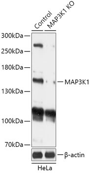
Western blot analysis of lysates from wild type (WT) and MAP3K1 knockout (KO) HeLa cells, using [KO Validated] MAP3K1 Rabbit pAb (CAB18041) at 1:1000 dilution. Secondary antibody: HRP-conjugated Goat anti-Rabbit IgG (H+L) (AS014) at 1:10000 dilution. Lysates/proteins: 25μg per lane. Blocking buffer: 3% nonfat dry milk in TBST. Detection: ECL Basic Kit (AbGn00020). Exposure time: 1min.
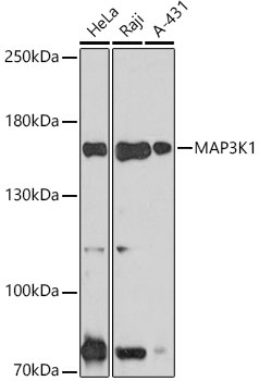
Western blot analysis of various lysates using MAP3K1 (CAB18041) at 1:1000 dilution. Secondary antibody: HRP-conjugated Goat anti-Rabbit IgG (H+L) (AS014) at 1:10000 dilution. Lysates/proteins: 25μg per lane. Blocking buffer: 3% nonfat dry milk in TBST. Detection: ECL Basic Kit (AbGn00020). Exposure time: 30s.
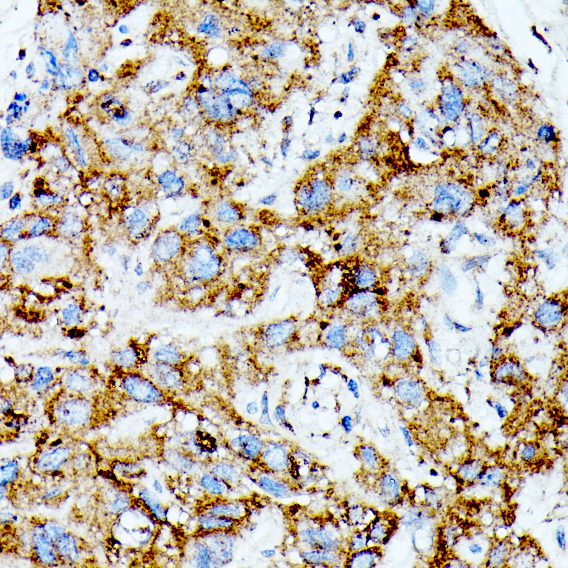
Immunohistochemistry analysis of paraffin-embedded Human liver cancer tissue using MAP3K1 Rabbit pAb (CAB18041) at a dilution of 1:100 (40x lens). Microwave antigen retrieval was performed with 0.01 M Tris-EDTA repair solution (pH 9.0) prior to IHC staining.
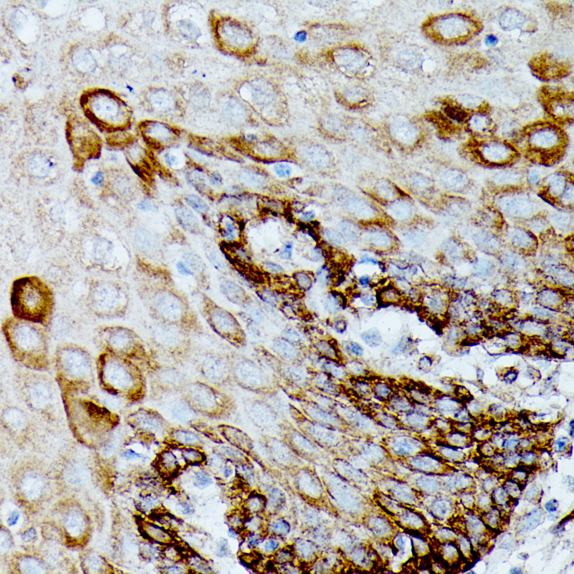
Immunohistochemistry analysis of paraffin-embedded Human esophagus tissue using MAP3K1 Rabbit pAb (CAB18041) at a dilution of 1:100 (40x lens). Microwave antigen retrieval was performed with 0.01 M Tris-EDTA repair solution (pH 9.0) prior to IHC staining.
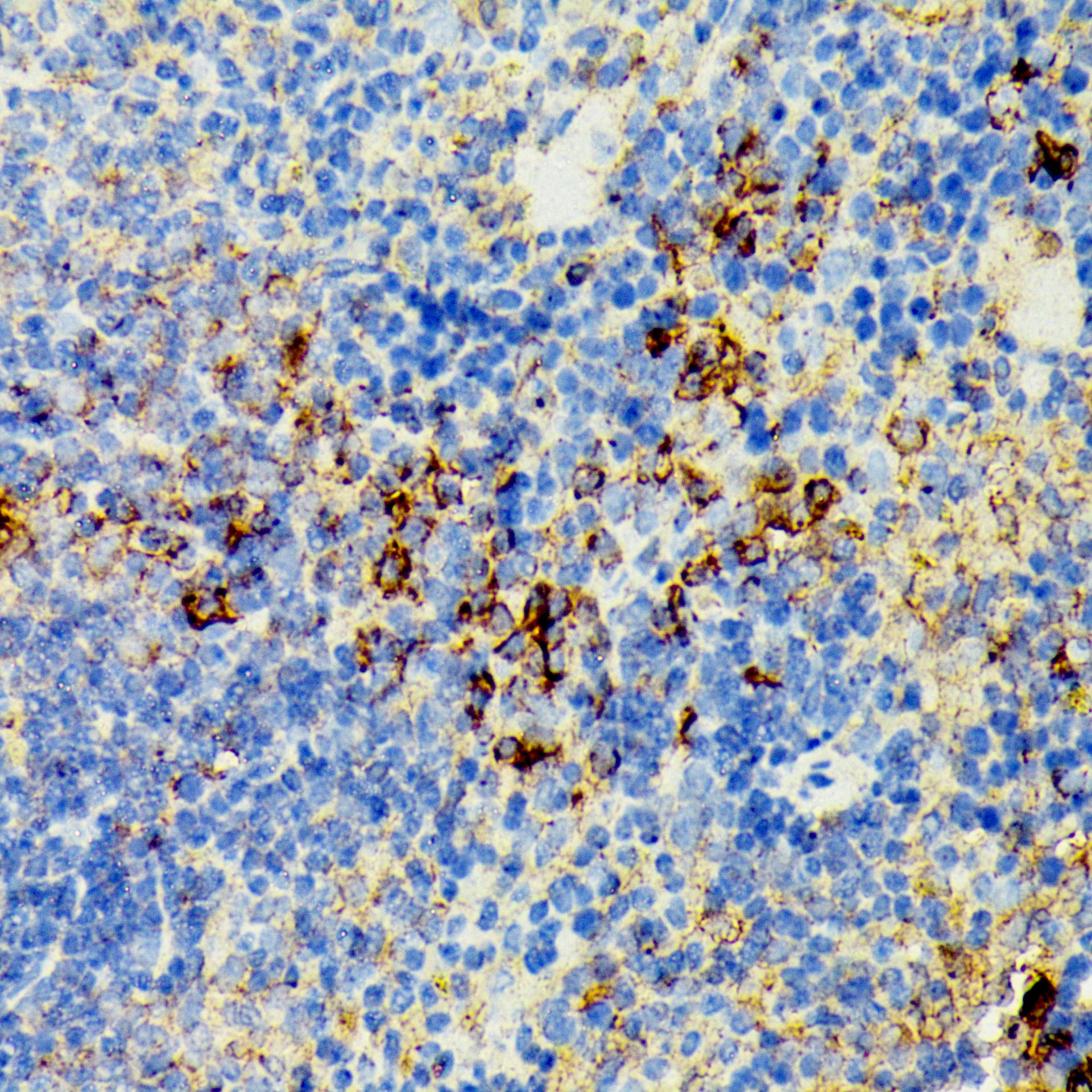
Immunohistochemistry analysis of paraffin-embedded Mouse spleen tissue using MAP3K1 Rabbit pAb (CAB18041) at a dilution of 1:100 (40x lens). Microwave antigen retrieval was performed with 0.01 M Tris-EDTA repair solution (pH 9.0) prior to IHC staining.
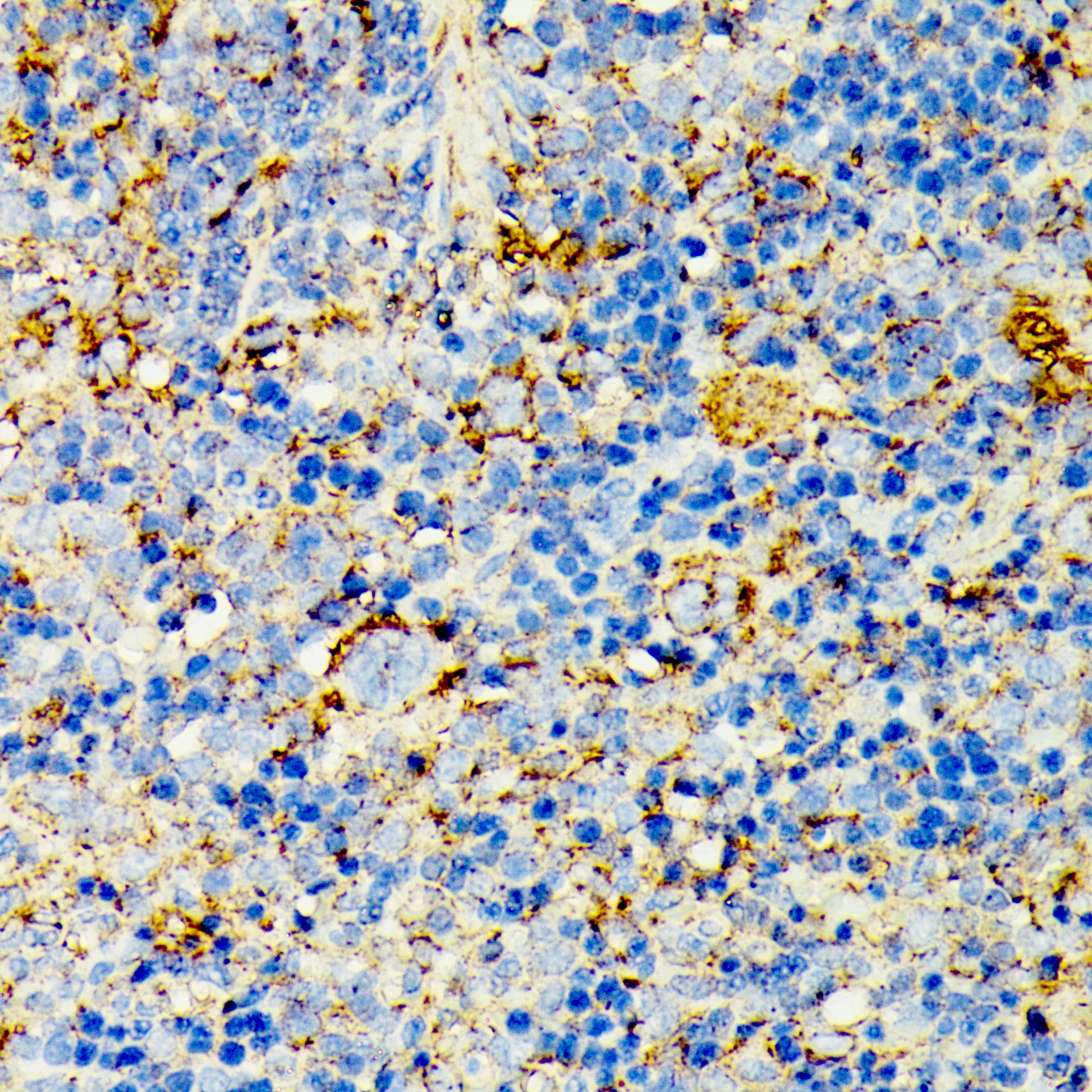
Immunohistochemistry analysis of paraffin-embedded Rat spleen tissue using MAP3K1 Rabbit pAb (CAB18041) at a dilution of 1:100 (40x lens). Microwave antigen retrieval was performed with 0.01 M Tris-EDTA repair solution (pH 9.0) prior to IHC staining.
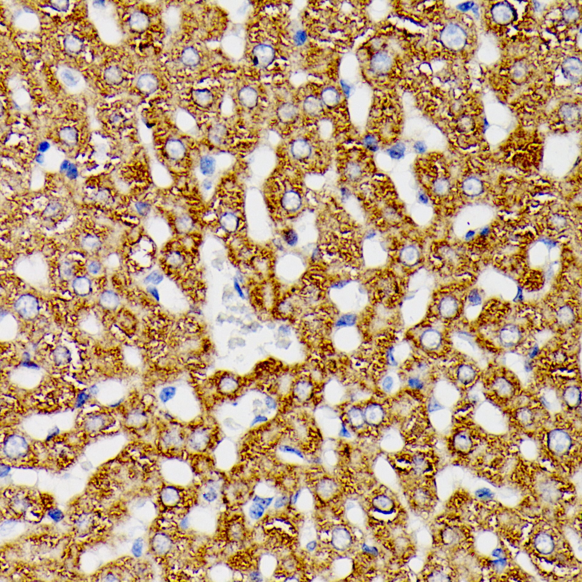
Immunohistochemistry analysis of paraffin-embedded Rat liver tissue using MAP3K1 Rabbit pAb (CAB18041) at a dilution of 1:100 (40x lens). Microwave antigen retrieval was performed with 0.01 M Tris-EDTA repair solution (pH 9.0) prior to IHC staining.