The [KO Validated] Smad5 Monoclonal Antibody (CAB19117) is a high-quality antibody developed for reliable detection and analysis of target proteins. This antibody, produced using rabbit-derived monoclonal technology, exhibits high specificity and sensitivity towards human samples, making it an invaluable reagent for Western blot applications. By targeting the SMAD5 protein, this antibody enables precise detection and analysis in a wide range of cell types, making it an ideal choice for studies in developmental biology and cancer research.SMAD5 is a vital mediator of the TGF-β signaling cascade, exerting control over a multitude of cellular processes including proliferation, migration, and apoptosis.
This antibody is validated for use in WB, IP, ELISA applications and has demonstrated reactivity against Human, Mouse samples.
Product Name:
[KO Validated] Smad5 Monoclonal Antibody
SKU:
CAB19117
Size:
20μL, 100μL
Reactivity:
Human, Mouse
Clone Number:
ARC0448
Conjugate:
Unconjugated
Immunogen:
Synthetic peptide. This information is considered to be commercially sensitive.
0.5μg-4μg antibody for 200μg-400μg extracts of whole cells
ELISA
Recommended starting concentration is 1 μg/mL. Please optimize the concentration based on your specific assay requirements.
Synonyms:
DWFC, JV5-1, MADH5, Smad5
Positive Sample:
K-562, C2C12
Cellular Localization:
Cytoplasm, Nucleus.
Calculated MW:
52kDa
Observed MW:
60kDa
The protein encoded by this gene is involved in the transforming growth factor beta signaling pathway that results in an inhibition of the proliferation of hematopoietic progenitor cells. The encoded protein is activated by bone morphogenetic proteins type 1 receptor kinase, and may be involved in cancer. Alternative splicing results in multiple transcript variants.
Purification Method
Affinity purification
Gene ID
4090
RRID
AB_2862610
Buffer Information
Store at -20℃. Avoid freeze / thaw cycles. Buffer: PBS containing 50% glycerol and 0.05% BSA, preserved with proclin300 or sodium azide, pH 7.3.
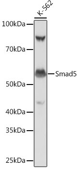
Western blot analysis of lysates from K-562 cells, using Smad5 Rabbit mAb (CAB19117) at 1:1000 dilution. Secondary antibody: HRP-conjugated Goat anti-Rabbit IgG (H+L) (CABS014) at 1:10000 dilution. Lysates/proteins: 25μg per lane. Blocking buffer: 3% nonfat dry milk in TBST. Detection: ECL Basic Kit (AbGn00020). Exposure time: 1s.
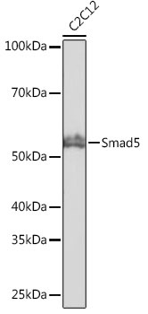
Western blot analysis of lysates from C2C12 cells, using Smad5 Rabbit mAb (CAB19117) at 1:1000 dilution. Secondary antibody: HRP-conjugated Goat anti-Rabbit IgG (H+L) (CABS014) at 1:10000 dilution. Lysates/proteins: 25μg per lane. Blocking buffer: 3% nonfat dry milk in TBST. Detection: ECL Basic Kit (AbGn00020). Exposure time: 10s.