The KPNA1 Antibody (CAB1742) is a high-quality antibody developed for reliable detection and analysis of target proteins. This antibody, produced in rabbits, is highly specific to human samples and has been validated for use in Western blot applications. By binding to KPNA1, this antibody allows for the detection and analysis of the protein in various cell types, making it ideal for studies in molecular biology and cancer research.KPNA1, also known as importin alpha-5, plays a crucial role in nucleocytoplasmic transport, enabling the movement of proteins into the nucleus for various cellular processes.
This antibody is validated for use in WB, IHC-P, ELISA applications and has demonstrated reactivity against Human, Mouse, Rat samples.
Product Name:
KPNA1 Antibody
SKU:
CAB1742
Size:
20μL, 100μL
Reactivity:
Human, Mouse, Rat
Conjugate:
Unconjugated
Immunogen:
Recombinant protein (or fragment).This information is considered to be commercially sensitive.
Recommended starting concentration is 1 μg/mL. Please optimize the concentration based on your specific assay requirements.
Synonyms:
RCH2, SRP1, IPOA5, NPI-1, KPNA1
Positive Sample:
Hep G2, 293T, Mouse brain
Cellular Localization:
Cytoplasm, Nucleus.
Calculated MW:
60kDa
Observed MW:
62kDa
The transport of molecules between the nucleus and the cytoplasm in eukaryotic cells is mediated by the nuclear pore complex (NPC), which consists of 60-100 proteins. Small molecules (up to 70 kD) can pass through the nuclear pore by nonselective diffusion while larger molecules are transported by an active process. The protein encoded by this gene belongs to the importin alpha family, and is involved in nuclear protein import. This protein interacts with the recombination activating gene 1 (RAG1) protein and is a putative substrate of the RAG1 ubiquitin ligase. Alternative splicing results in multiple transcript variants.
Purification Method
Affinity purification
Gene ID
3836
RRID
AB_2763788
Buffer Information
Store at -20℃. Avoid freeze / thaw cycles. Buffer: PBS containing 50% glycerol, preserved with proclin300 or sodium azide, pH 7.3.
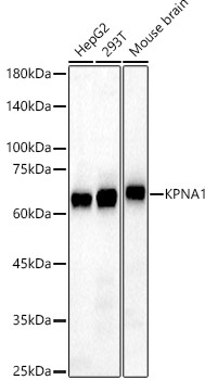
Western blot analysis of various lysates, using KPNA1 Rabbit pAb (CAB1742) at 1:1000 dilution. Secondary antibody: HRP-conjugated Goat anti-Rabbit IgG (H+L) (CABS014) at 1:10000 dilution. Lysates/proteins: 25μg per lane. Blocking buffer: 3% nonfat dry milk in TBST. Detection: ECL Basic Kit (AbGn00020). Exposure time: 10s.
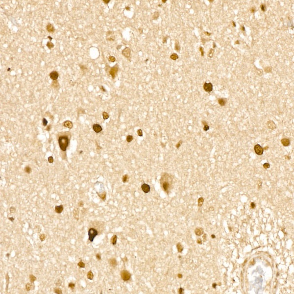
Immunohistochemistry analysis of paraffin-embedded Human brain tissue using KPNA1 Rabbit pAb (CAB1742) at a dilution of 1:100 (40x lens). High pressure antigen retrieval was performed with 0.01 M citrate buffer (pH 6.0) prior to IHC staining.
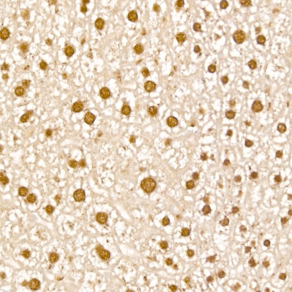
Immunohistochemistry analysis of paraffin-embedded Mouse liver tissue using KPNA1 Rabbit pAb (CAB1742) at a dilution of 1:100 (40x lens). High pressure antigen retrieval was performed with 0.01 M citrate buffer (pH 6.0) prior to IHC staining.
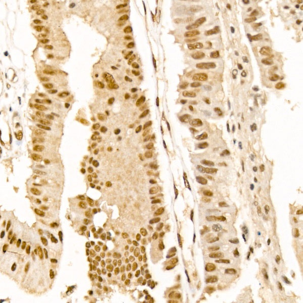
Immunohistochemistry analysis of paraffin-embedded Human colon carcinoma tissue using KPNA1 Rabbit pAb (CAB1742) at a dilution of 1:100 (40x lens). High pressure antigen retrieval was performed with 0.01 M citrate buffer (pH 6.0) prior to IHC staining.